Tetralogy of Fallot (TOF) is the most common cyanotic congenital heart disease beyond the infantile period, accounting for approximately 12% of all congenital heart defects. The condition was named after the French physician Etienne Fallot, who, in 1888, provided a detailed description of its pathological changes and clinical manifestations. Approximately 25% of cases involve a right-sided aortic arch. Additional cardiovascular abnormalities, such as persistent left superior vena cava, coronary artery anomalies, atrial septal defect, patent ductus arteriosus, or absent pulmonary valve, may also coexist.
Pathological Anatomy
Tetralogy of Fallot consists of four specific abnormalities. Among these, right ventricular outflow tract obstruction is the primary determinant of the pathophysiology, severity of the condition, and prognosis. This obstruction tends to worsen progressively over time.
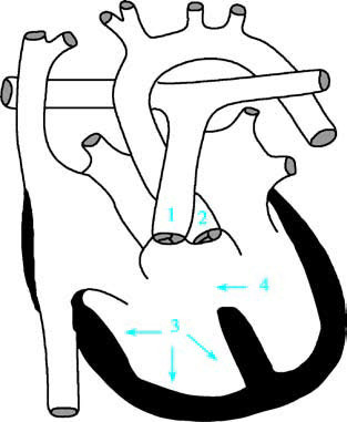
Figure 1 Diagram of tetralogy of Fallot
1, Right ventricular infundibular and pulmonary valve stenosis;
2, Overriding aorta;
3, Right ventricular hypertrophy;
4, Ventricular septal defect.
Right Ventricular Outflow Tract Obstruction
The obstruction can extend from the entrance of the right ventricular infundibulum to the branches of the left and right pulmonary arteries. It may involve infundibular stenosis, valvular stenosis, or a combination of both. Pulmonary valve ring, the main pulmonary artery, and pulmonary artery branches may exhibit underdevelopment or asymmetric narrowing. The severity of the obstruction can vary widely.
Ventricular Septal Defect
The defect is typically perimembranous and extends toward the outflow tract, usually positioned beneath the aorta, and may extend toward the pulmonary artery. It is considered a malalignment type of ventricular septal defect.
Overriding Aorta
The aortic root is enlarged, rotating clockwise and shifting toward the right, straddling the ventricular septal defect. The degree of overriding can range from 15% to 95%.
Right Ventricular Hypertrophy
This is generally regarded as a secondary feature, resulting from compensatory changes caused by increased workload on the right ventricle.
Pathophysiology
The ventricular septal defect in Tetralogy of Fallot is nonrestrictive, resulting in equal pressure in both the left and right ventricles. Depending on the severity of the right ventricular outflow tract obstruction, the direction of blood shunting at the ventricular level may be left-to-right, bidirectional, or right-to-left.
In cases of mild pulmonary artery stenosis, left-to-right shunting can occur, leading to minimal or absent cyanosis. Severe pulmonary artery stenosis causes significant right-to-left shunting and notable clinical cyanosis. The murmur originates from the right ventricular outflow tract obstruction, not from the ventricular septal defect. The obstruction leads to increased afterload on the right ventricle, triggering compensatory hypertrophy.
The overriding aorta, positioned above both the right and left ventricles, directly receives venous blood from the right ventricle in addition to the oxygenated blood from the left ventricle. This results in systemic cyanosis. Additionally, pulmonary artery stenosis reduces blood flow to the lungs for gas exchange, further exacerbating the cyanosis. Decreased pulmonary blood flow may also promote the formation of collateral circulation between the enlarged bronchial arteries and pulmonary vessels.
Before the closure of the ductus arteriosus, the reduction in pulmonary blood flow may be less pronounced, resulting in mild or absent cyanosis. As the ductus arteriosus closes and infundibular stenosis worsens, cyanosis becomes increasingly noticeable, often accompanied by the development of clubbing of the fingers and toes.
Chronic hypoxia stimulates compensatory overproduction of red blood cells in the bone marrow, leading to increased blood viscosity and sluggish circulation. This condition can result in cerebral thrombosis. If bacterial thrombi are present, there is a heightened risk of brain abscess formation.
Clinical Manifestations
Cyanosis
Cyanosis is the primary clinical feature of Tetralogy of Fallot. Its severity and the age of onset are associated with the degree of pulmonary artery stenosis and whether the ductus arteriosus is closed. Cyanosis often occurs in superficial areas rich in capillaries, such as the lips, nail beds, and bulbar conjunctiva. Reduced arterial oxygen saturation leads to poor exercise tolerance. Even mild activity, such as crying, emotional stress, physical exertion, or exposure to cold, may worsen dyspnea and intensify cyanosis.
Squatting
Squatting is a common compensatory behavior, especially during activities such as walking or playing. Children often squat for brief periods. Squatting flexes the lower limbs, reducing venous return to the heart and thereby alleviating cardiac workload. Compression of the lower arteries by the thighs increases systemic vascular resistance, reducing right-to-left shunting and temporarily mitigating hypoxia. Infants who cannot walk are often held in a sitting position with their legs flexed.
Clubbing of Fingers and Toes
Persistent cyanosis lasting more than six months can lead to clubbing of the fingers and toes. Chronic hypoxia causes capillary dilation and proliferation in the tips of the digits, along with soft tissue and bone hypertrophy, leading to the characteristic drumstick-like appearance.
Paroxysmal Hypoxic Spells
Paroxysmal hypoxic spells are most common in infants and may be triggered by factors such as feeding, crying, emotional agitation, anemia, or infection. These episodes manifest as sudden dyspnea and, in severe cases, syncope, seizures, or even death. The spells are thought to be caused by muscle spasms in the already stenotic infundibulum, leading to transient obstruction of the pulmonary artery and exacerbation of cerebral hypoxia. Older children may report headaches or dizziness during these episodes.
Growth and development in children with Tetralogy of Fallot are often delayed, and intellectual development may also lag slightly behind that of their peers. Physical examination may reveal a slight protrusion of the precordium. A grade 2–3 harsh, ejective systolic murmur is often audible at the left sternal border in the 2nd to 4th intercostal spaces, originating from the pulmonary artery stenosis. A systolic thrill is generally absent. The second pulmonary artery sound is typically diminished. In some patients, a single, accentuated second heart sound may be heard, originating from the overriding aorta. With severe stenosis or during hypoxic spells, murmurs may become inaudible. Continuous murmurs may occasionally be heard, indicative of collateral circulation.
Common complications include cerebral thrombosis, brain abscess, and infective endocarditis.
Auxiliary Examinations
Blood Tests
Peripheral red blood cell count and hemoglobin concentration are significantly elevated. Red blood cell counts may reach (5.0–8.0) × 1012/L, with hemoglobin levels ranging from 170 to 200 g/L. Hematocrit is also increased, ranging from 53% to 80%. Thrombocytopenia is observed, and prothrombin time is prolonged.
X-ray Examination
The heart size is generally normal or slightly enlarged. On a standard anteroposterior chest X-ray, a characteristic "boot-shaped" cardiac silhouette may be seen, with an upturned and blunt apex, concavity in the pulmonary artery segment, and widening of the upper mediastinum. Pulmonary hilar vasculature is reduced, and the transparency of the lung fields increases due to decreased vascular markings in both lungs. Older children may exhibit a reticular pattern in the lung fields due to the development of collateral circulation. A right-sided aortic arch is present in 25% of cases.
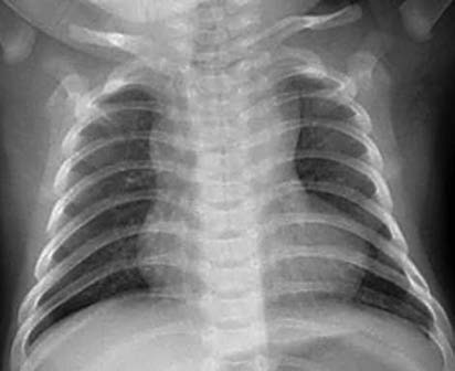
Figure 2 Chest X-Ray of tetralogy of Fallot (Anteroposterior view)
Electrocardiography (ECG) Examination
Findings include right axis deviation, right ventricular hypertrophy, and signs of myocardial strain in severe cases of obstruction. Enlargement of the right atrium may also be observed.
Echocardiography
Two-dimensional echocardiography reveals an enlarged aortic diameter overriding the ventricular septum, interruption of the ventricular septum, and variable degrees of aortic override and stenosis in the right ventricular outflow tract and pulmonary artery. Additionally, the diameters of the right ventricle and right atrium are increased, whereas the diameter of the left ventricle is reduced. Color Doppler imaging demonstrates blood flow directly from the right ventricle into the overriding aorta and allows visualization of shunting across the ventricular septal defect.
Cardiac Catheterization
Cardiac catheterization and angiography are indicated for patients with abnormalities such as underdeveloped peripheral pulmonary artery branches or systemic-to-pulmonary collateral circulation. Selective left ventricular and aortic angiography can help evaluate left ventricular development as well as the origin and course of the coronary arteries.
Treatment
Medical Management
General Care
Sufficient fluid intake should be ensured to prevent dehydration and associated complications. Infants and young children require special attention to prevent hypoxic episodes. Infection prevention is essential.
Management of Hypoxic Episodes
Mild episodes may resolve with the knee-chest position. Severe episodes often necessitate oxygen supplementation and administration of deoxyepinephrine at a dose of 0.05 mg/kg via intravenous injection or propranolol at 0.1 mg/kg per dose. Morphine injections (0.1–0.2 mg/kg subcutaneously) may also be administered if necessary. Acidosis should be corrected with 5% sodium bicarbonate (1.5–5.0 mL/kg intravenously). For those with a history of hypoxic episodes, oral propranolol (1–3 mg/kg/day) may be utilized. Triggers of hypoxic episodes, such as anemia or infection, should be addressed, and the child’s environment should remain as calm as possible. Emergency surgical repair should be considered for refractory hypoxic episodes.
Surgical Management
Surgery is considered the definitive treatment for tetralogy of Fallot. Patients with mild conditions may undergo one-stage corrective surgery during the preschool years, while those with significant clinical symptoms are typically operated on within the first six months of life. For severe cases, palliative surgery may be performed initially. Definitive repair is conducted after general health improves and pulmonary vasculature matures. Common palliative procedures include subclavian artery-to-pulmonary artery shunts (modified Blalock-Taussig shunt).
Prognosis
Surgical outcomes for tetralogy of Fallot have significantly improved in recent years, with declining operative mortality rates. Long-term prognosis is favorable for most patients. Long-term follow-up is required post-surgery to monitor for residual pulmonary obstruction, pulmonary regurgitation, residual ventricular septal shunts, arrhythmias, and cardiac dysfunction.