Fetal structural anomaly refers to significant structural or anatomical differences compared to a normal fetus. Common presentations include malformation, dysplasia, deformation, or disruption. The causes of fetal structural anomalies include genetic factors, environmental factors, or the interaction of both.
Screening for fetal structural anomalies is primarily performed through ultrasound examinations between 20 and 24 weeks of gestation. The goal of prenatal ultrasound screening is to detect severe, life-threatening, or disabling structural anomalies, such as anencephaly, spina bifida, holoprosencephaly, encephalocele, a single ventricle with a single great artery, gastroschisis, lethal dwarfism, and bilateral renal agenesis.
Anencephaly
Anencephaly is one of the most common severe birth defects, caused by the failure of the anterior neuropore to close. It represents the most severe form of neural tube defects. Female fetuses are four times more likely to be affected than males. Due to the absence of cranial bones, the eyeballs protrude, giving a "frog-like" facial appearance. The neck is shortened, and the brain is absent, with only the cranial base or partial brain tissue present, making survival impossible. Anencephaly can be detected with high sensitivity during prenatal ultrasound screening. Beyond 14 weeks of gestation, ultrasound may reveal the absence of the rounded cranial bone outline and irregular "nodule-like" structures at the cephalic end. Anencephaly is a severe and fatal birth defect. Once diagnosed, termination of pregnancy is recommended.
Spina Bifida
Spina bifida is a condition in which parts of the spinal canal fail to completely close. It is the most common type of neural tube defect, with its incidence varying significantly by region and ethnicity. Spinal ossification begins around 8 to 9 weeks of gestation, and failure of the vertebral halves to fuse results in spina bifida, which most commonly affects the thoracolumbar region.
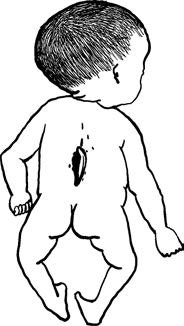
Figure 1 Spina bifida
There are three main types of spina bifida:
- Occult Spina Bifida: Characterized by a defect in the spinal canal, often located at the lumbosacral region and covered by skin. The spinal cord and spinal nerves are generally normal, with no neurological symptoms.
- Meningomyelocele: This involves a defect in two vertebrae, with herniation of the meninges through the spinal defect. Larger herniations may include meninges, spinal cord, and nerve roots. This form is often associated with neurological symptoms.
- Myeloschisis: A more severe condition where there is a failure in neural tube formation, leaving the neural tissue open and exposed.
Occult spina bifida is often difficult to detect during prenatal ultrasound. However, open spina bifida is more easily identified through prenatal ultrasound, with the 18–20 week gestation period being the optimal timing. Ultrasound may reveal a widened space between two rows of hyperechoic spinal reflections or abnormal angular patterns resembling "V" or "W" shapes, along with shortened, irregularly curved spines or cystic protrusions. Pregnant individuals at high risk based on maternal serum AFP screening or ultrasound findings are advised to undergo targeted ultrasound evaluations during the mid-pregnancy period.
The prognosis for spina bifida varies. Once diagnosed prenatally, decisions regarding pregnancy management should consider gestational age at diagnosis, severity, and the preferences of the pregnant individual and their family. For those who opt to continue the pregnancy, a multidisciplinary approach involving obstetrics, neonatology, and pediatric neurosurgery is essential to create an integrated prenatal and postnatal management plan, aiming to improve survival rates and reduce long-term neurological complications.
Holoprosencephaly
Holoprosencephaly, also referred to as the failure of prosencephalon division, is a brain anomaly resulting from the embryonic forebrain failing to divide into two distinct cerebral hemispheres during the third to fourth weeks of gestation. Holoprosencephaly has three classic types: alobar, semilobar, and lobar.
Alobar holoprosencephaly is the most severe form and is characterized by the absence of the longitudinal fissure and falx cerebri, presenting only a single primitive ventricle. The thalami are fused at the midline, and facial abnormalities are often present. Holoprosencephaly is a severe central nervous system malformation frequently associated with facial anomalies and genetic disorders, such as trisomy 13. Alobar holoprosencephaly is considered a lethal structural anomaly, and termination of pregnancy is recommended following diagnosis.
Encephalocele
Encephalocele refers to brain tissue and meninges herniating through cranial bone defects. It commonly occurs along the midline, most frequently at the occipital region, followed by the frontal region. Severe encephaloceles may be detected during early pregnancy ultrasound. Ultrasound findings include an incomplete skull outline, with a mixed echogenic mass protruding through the cranial defect. The extruding brain tissue is often connected to the intracranial parenchyma and may be accompanied by ventriculomegaly.
The causes of encephalocele include chromosomal abnormalities, such as trisomy 13 and trisomy 18, as well as single-gene disorders like Meckel-Gruber syndrome and Walker-Warburg syndrome. Severe encephalocele is considered a fatal anomaly. Once diagnosed, termination of pregnancy is typically recommended.
Single Ventricle and Single Great Artery
Single ventricle refers to a condition where a single ventricle receives blood from both atria, or from a common atrium, with or without a residual small chamber. It is associated with atrioventricular valves that are either distinctly left and right or fused into a common atrioventricular valve. This represents a severe congenital cardiac malformation with poor prognosis. On ultrasound imaging, the four-chamber view typically shows only one main ventricular chamber or a single large primary chamber with a smaller residual chamber.
A single great artery refers to the presence of only one main arterial trunk, with the absence of or failure to visualize the second main arterial trunk. Common types include truncus arteriosus, pulmonary atresia, and aortic atresia.
Although advances in pediatric cardiac surgery have significantly improved survival rates for various procedures, most treatments still require staged surgeries. Postoperative care demands substantial social and medical resources. Therefore, thorough communication between medical professionals, the pregnant individual, and their family is essential. Decision-making regarding the pregnancy is advised to be guided by a multidisciplinary consultation involving fetal medicine specialists and pediatric cardiologists.
Gastroschisis
Gastroschisis is caused by a full-thickness defect in one side of the anterior abdominal wall. Prenatal ultrasound typically reveals a discontinuity in the echo of the fetal abdominal wall, an empty fetal abdominal cavity, and intra-abdominal organs such as the stomach and intestines floating freely in the amniotic fluid without any covering membrane.
With advancements in pediatric surgical techniques, the survival rate for isolated gastroschisis caused by non-genetic factors and not associated with other structural anomalies exceeds 90%. However, cases involving liver herniation are associated with increased mortality rates. For pregnancies continuing with a diagnosis of gastroschisis, multidisciplinary consultation involving fetal medicine specialists, geneticists, pediatric surgeons, and obstetricians is advised to formulate an integrated management strategy for both the prenatal and postnatal periods. This should include evaluation of the feasibility of surgical intervention immediately after delivery, assessment of any associated anomalies, and timely referrals for early surgical treatment.
Thanatophoric Dwarfism
Thanatophoric dwarfism is the most common lethal condition associated with skeletal dysplasia. It is characterized by extremely short and curved long bones, a narrowed thoracic cavity, a disproportionately large head, and an enlarged abdomen, often accompanied by polyhydramnios. On ultrasound imaging, fetal long bones appear "telephone receiver"-shaped, with shortened and curved femurs and humeri being especially notable.
The fatal outcomes of this condition are primarily due to extreme thoracic narrowing, which leads to pulmonary hypoplasia and cardiopulmonary failure. It has been established that thanatophoric dwarfism is caused by mutations in the FGFR3 gene, with definitive diagnosis relying on genetic testing. This condition is sporadic and has an extremely low recurrence risk. Upon identification of thanatophoric dwarfism, pregnancy termination is recommended as early as possible.
Bilateral Renal Agenesis
Bilateral renal agenesis occurs due to the failure of both ureteric buds to develop, preventing the differentiation of the metanephric mesenchyme into functional kidneys. Prenatal diagnosis primarily relies on ultrasound findings, which include the absence of renal tissue on both sides of the renal fossa and surrounding areas, flattened adrenal glands (lying-flat sign), progressive oligohydramnios during the second trimester, and eventual anhydramnios. Severe oligohydramnios leads to pulmonary hypoplasia. When poor image quality caused by minimal amniotic fluid complicates ultrasound diagnosis, fetal MRI may provide additional diagnostic support.
Bilateral renal agenesis is considered a lethal structural anomaly. Once diagnosed, termination of pregnancy is typically recommended.