Early pregnancy, also referred to as early gestation, is a crucial period for embryo formation and fetal organ differentiation. The primary goals of early diagnosis are to confirm pregnancy, determine the number of fetuses, estimate gestational age, and rule out pathological conditions such as ectopic pregnancy.
Symptoms and Signs
Missed Menstruation
Healthy women of reproductive age with a history of sexual activity and regular menstrual cycles should consider the possibility of pregnancy if menstruation is delayed. A delay of more than 10 days raises a strong suspicion of pregnancy. While a missed period is the earliest symptom of pregnancy, it is not exclusive to pregnancy, as some individuals may experience irregular vaginal bleeding after conception without a clear history of missed menstruation.
Early Pregnancy Symptoms
Around six weeks after the last menstrual period, symptoms such as chills, dizziness, excessive salivation, fatigue, drowsiness, poor appetite, a preference for sour foods, aversion to greasy foods, nausea, and morning vomiting may occur. These are collectively termed early pregnancy symptoms. Some individuals may also experience mood changes. Symptoms typically resolve naturally by approximately 12 weeks of gestation.
Frequent Urination
Frequent urination results from the forward-tilting and enlargement of the uterus, which presses on the bladder in the pelvis. This symptom usually resolves once the uterus grows beyond the pelvic cavity.
Breast Changes
Breast tenderness and enlargement are common sensations. On examination, breasts gradually increase in size with prominent visible veins. The nipples enlarge, and both the nipples and areolae darken in color. Enlarged sebaceous glands around the areola, known as Montgomery's tubercles, become visible as deep brown nodules. In lactating women, milk production significantly decreases after conception.
Gynecological Examination
The vaginal mucosa and vaginal portion of the cervix exhibit congestion with a bluish-purple coloration. The cervical isthmus becomes extremely soft. During a bimanual examination at 6–8 weeks of gestation, a separation-like sensation between the cervix and uterine body, known as Hegar's sign, may be felt. The uterus gradually enlarges, softens, and becomes more spherical. By 8 weeks of pregnancy, the size of the uterus doubles compared to that of the non-pregnant state; by 12 weeks, it triples in size, with the uterine fundus palpable above the pubic symphysis.
Other Symptoms
Some individuals present with signs of increased estrogen levels, such as spider angiomas, palmar erythema, and skin hyperpigmentation on the face, linea alba, and areolae.
Auxiliary Examinations
Pregnancy Test
Human chorionic gonadotropin (hCG) levels in the serum can rise shortly after implantation of the fertilized egg, as detected by radioimmunoassay. Clinically, urine pregnancy tests using early pregnancy test kits are common. A positive result combined with clinical symptoms indicates pregnancy. However, ultrasound examination is necessary to confirm intrauterine pregnancy.
Ultrasound Examination
The primary objectives of early pregnancy ultrasound are to confirm intrauterine pregnancy, rule out ectopic pregnancy and gestational trophoblastic disease, estimate gestational age, and exclude pelvic masses or uterine anomalies. In cases of multiple pregnancies, the number and morphology of gestational sacs can determine chorionicity. Around 35 days after the last menstrual period, a round or oval-shaped gestational sac (GS) may be seen in the uterine cavity. By 6 weeks, the embryo and primitive cardiac activity become visible. Before 14 weeks of pregnancy, the crown-rump length (CRL) is an accurate measurement to estimate gestational age and adjust the estimated due date. Between 11 and 13 weeks and 6 days, ultrasound can identify severe fetal malformations, such as anencephaly. Ultrasound parameters, including nuchal translucency (NT) thickness and nasal bone presence, are useful for chromosomal abnormality screening during the first trimester. Color Doppler ultrasound can detect fetal cardiac activity to confirm early pregnancy and fetal viability.
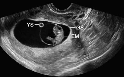
Figure 1 Early pregnancy ultrasound image
GS: Gestational sac
EM: Embryo
YS: Yolk sac
Diagnosis
Pregnancy should be considered in reproductive-aged women with a history of sexual activity within the current menstrual cycle who experience missed periods or abnormal vaginal bleeding. Positive blood or urine hCG levels support the diagnosis of pregnancy. The identification of an intrauterine gestational sac and embryo on ultrasound confirms intrauterine pregnancy, and the presence of cardiac activity indicates a viable embryo. A definitive diagnosis of normal pregnancy requires the combination of blood or urine hCG positivity, ultrasound identification of the embryo, and detection of cardiac activity.