The normal development of female reproductive organs is a highly complex process. The genetic sex of humans is determined at fertilization by the karyotype of the zygote. However, the sex of the gonads can only be identified by the 7th week of embryonic development, and the sex of the external genitalia becomes distinguishable by the 12th week of fetal development. Female embryonic cells have XX sex chromosomes, and the undifferentiated gonads develop into ovaries. The Müllerian duct, also known as the paramesonephric duct, serves as the precursor of the female reproductive tract. In the absence of androgen, the external genitalia naturally differentiate into female structures.
Development of the Gonads
The gonads, or sex glands, develop from the gonadal ridge, which consists of coelomic epithelium, underlying mesenchyme, and migrating primordial germ cells. During the 3rd to 4th week of embryonic development, primordial germ cells, which are larger than surrounding somatic cells, appear in the endoderm of the yolk sac. By the 5th week of development, the proliferation of coelomic epithelium on the medial side of the mesonephros results in the formation of the urogenital ridge, with the lateral elevation forming the mesonephric ridge and the medial elevation forming the gonadal ridge. Proliferating epithelial cells on the gonadal ridge form the gonadal primordium. These epithelial cells extend into the mesenchyme below, forming irregular cell cords known as primary sex cords. Before the 7th week of embryonic development, the gonads remain undifferentiated in both sexes and are referred to as indifferent gonads, appearing identical in male and female individuals.
After the 10th week of embryonic development, during the differentiation of the gonads into ovaries, the primary sex cords degenerate and are replaced by stroma and blood vessels, forming the ovarian medulla. Proliferating epithelial cells on the surface of the indifferent gonad extend again into the mesenchyme to form secondary sex cords. After separating from the epithelium, these secondary sex cords constitute the ovarian cortex. The mesenchyme beneath the epithelium forms the tunica albuginea.
The development of the gonads is influenced by the genotype and sex chromosomes of the fetus, while the ultimate phenotypic sex is determined by sex chromosomes as well as the predominant biochemical and hormonal environment. Under the influence of two X chromosomes, the cortex of the indifferent gonads tends to differentiate into female gonads. In male fetuses, the sex-determining region of the Y chromosome (SRY) encodes the testis-determining factor (TDF), which induces differentiation of the indifferent gonads into testes and the production of androgens. In addition to TDF and androgens, anti-Müllerian hormone (AMH) is critical for male development. In the absence of these three factors, the genitalia develop in a female direction. Subsequently, the maturation of female reproductive organs is primarily influenced by estrogen.
Development of Female Reproductive Tracts
Development of the Fallopian Tubes, Uterus, Cervix, and Upper Vagina
During the 7th week of embryonic development, the mesonephric duct degenerates, and the two Müllerian ducts continue to grow and extend caudally. The non-fused cranial portions of the two Müllerian ducts remain tubular and develop into the fallopian tubes, with their cranial ends forming the fimbriae. The middle and caudal portions of the Müllerian ducts fuse to form the uterus, cervix, and the upper one-third of the vagina. During the initial stages of fusion, a longitudinal septum exists within the uterine cavity. This septum is typically absorbed by the 20th week of fetal development, resulting in a single cavity.
Development of the Lower Reproductive Tract
During the 3rd week of embryonic development, the cloacal membrane forms below the umbilical cord. In the 4th week, the cloacal folds fuse anteriorly to form the genital tubercle. By the 7th week, the urorectal septum merges with the cloacal membrane, separating the rectum from the urogenital tract. A canal forms in the urogenital membrane, communicating with the amniotic cavity, and this canal develops into the primitive urogenital sinus. The primitive urogenital sinus eventually differentiates into a pelvic portion and an external portion. In females, the caudal pelvic portion of the urogenital sinus forms the urethra and the lower two-thirds of the vagina.
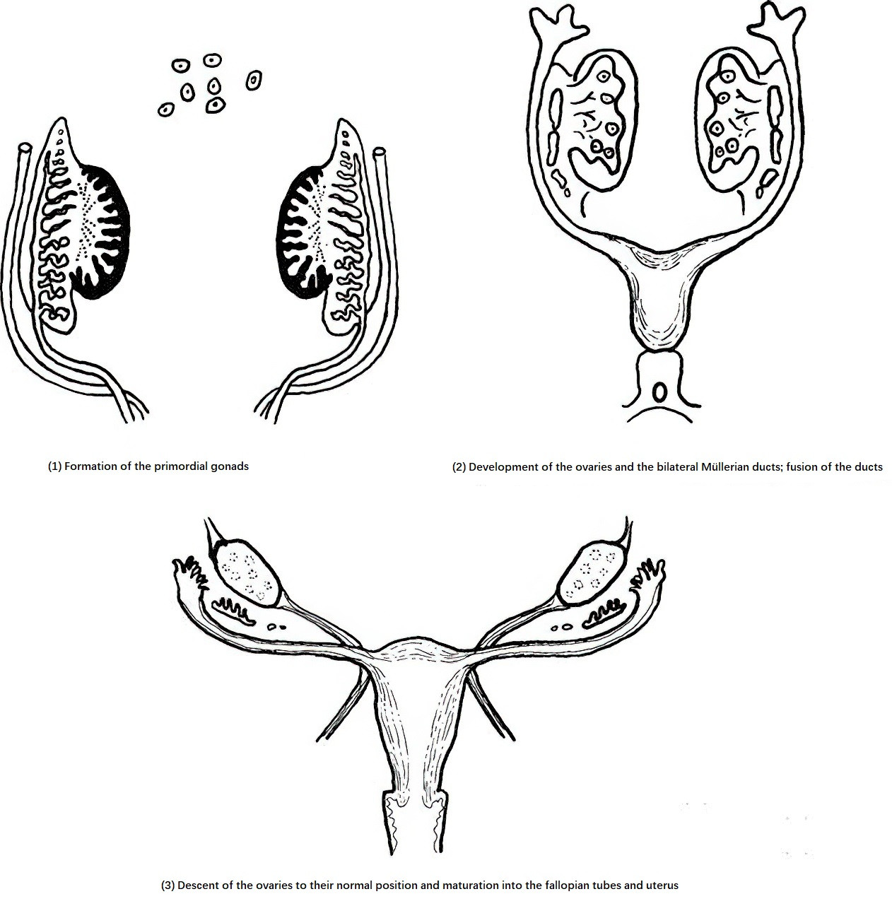
Figure 1 Ovary and internal reproductive development
Development of Female External Genitalia
By the 6th week of embryonic development, parts of the cloacal membrane invaginate locally to form the urethral and anal depressions. Surrounding the primitive urogenital groove, the urethral folds form medially, and the labioscrotal swellings form laterally. By the 7th week, the cloacal membrane disappears, establishing communication between the primitive urogenital groove and the urogenital sinus.
By the 10th week, sexual dimorphism in the external genitalia begins to appear, and by the 12th week, the differentiation is nearly complete. The unfused labioscrotal swellings develop into the labia majora, while the fused anterior portions form the mons pubis and the anterior commissure of the labia. The unfused urethral folds develop into the labia minora, and the posterior portions of these folds fuse to form the frenulum of the labia. The urogenital groove expands and, along with the caudal portion of the urogenital sinus, forms the vestibule of the vagina. By the 14th week of embryonic development, the genital tubercle develops into the clitoris.