Obtaining a detailed medical history prior to the examination is beneficial for accuracy. During the examination, the patient is positioned in a sitting posture with the entire neck and upper chest fully exposed. The process is carried out sequentially under adequate lighting and includes inspection, palpation, auscultation, and transillumination testing.
Inspection
The overall appearance of the neck is observed, noting any restrictions in movement or the presence of torticollis or forced head positioning. Symmetry between the sides of the neck is evaluated, along with the presence of any swelling or masses. Signs of venous distension and abnormal vascular pulsations are assessed. Attention is given to the position and shape of the laryngeal prominence, noting any laryngeal enlargement. The skin is checked for redness, swelling, ulcers, rashes, fistulae, or scars. Enlargement of the parotid gland, submandibular gland, and thyroid gland is noted.
Palpation
Palpation is performed while the patient remains fully relaxed. Assessment includes the range of motion of the neck in forward, backward, and lateral directions. Each region of the neck is palpated in sequence:
Submental and Submandibular Regions
The patient remains seated while the examiner stands facing the patient. One hand is placed on the occipital area of the patient’s head to assist in head movement, while the palmar surface of the fingers of the other hand palpates the submental and submandibular regions. Enlarged lymph nodes or swelling of the submandibular gland is noted.
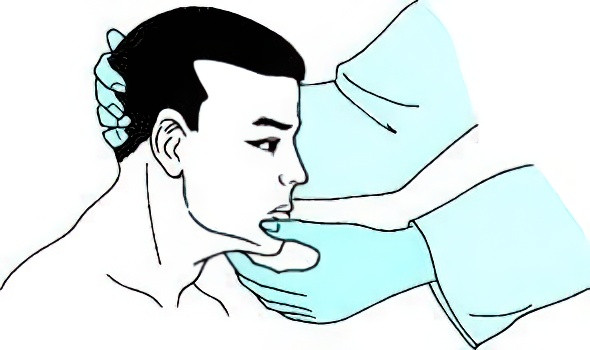
Figure 1 Examination for the submental and submandibular regions
Anterior Cervical Region
The thyroid gland is palpated first. Commonly, the examination is conducted with the patient seated while the examiner stands behind them. Both thumbs are placed on the back of the patient’s neck, and the index and middle fingers of each hand palpate the lateral lobes of the thyroid gland on each side. The size, shape, texture, presence of masses, tenderness, and whether the masses move with swallowing are noted. The trachea is also assessed for displacement or softening. In suspected cases of thyroglossal duct cyst, the cyst is palpated using the thumb and index finger, while the patient is asked to extend their tongue or swallow to observe cyst movement. Where laryngeal cancer is suspected, particularly if involvement of the laryngeal structure is suspected, the examiner lifts the larynx gently using the thumb and middle finger, moving it side-to-side to assess for enlargement or restricted movement. Lymph nodes in the pre-epiglottic space, anterior to the larynx, and anterior to the trachea are palpated for size, texture, mobility, and whether they are singular or clustered. The examiner may also perform the examination from a seated or standing position in front of the patient.
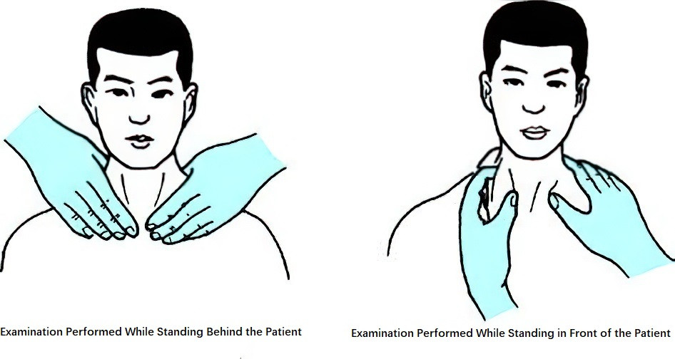
Figure 2 Examination for the anterior cervical region
Lateral Cervical and Supraclavicular Regions
The examiner stands facing the patient, placing one hand on the patient’s occipital area to assist neck rotation, while the other hand palpates the lateral cervical region deep to the sternocleidomastoid muscle. For the posterior triangle, the patient’s head is rotated toward the side being examined and slightly tilted backward. Supraclavicular regions are assessed with the examiner positioned behind the patient, using the thumb to press on the patient’s shoulder while the remaining fingers palpate the supraclavicular fossa. Swelling or masses in the neck are noted, along with their size, texture, mobility, singularity, or multiplicity, and whether they are discrete or fused. Associated tenderness or pulsations, as well as the presence of skin fistulae, are also observed. If a fistula is detected, its depth and direction can be examined using finger palpation or the insertion of a probe.
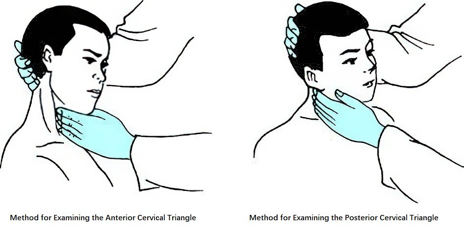
Figure 3 Examination for the lateral cervical region
Auscultation
In patients with hyperthyroidism, increased blood flow within the thyroid gland can produce a continuous systolic murmur detectable in the thyroid region. Patients with carotid body tumors may present with clear vascular murmurs in the carotid triangle. In carotid artery aneurysms, systolic murmurs may be heard at the sites of the arterial swelling. In cases of pharyngeal or cervical esophageal diverticulum, air sounds may be audible in the neck during swallowing. In cases of laryngeal obstruction, stridor may also be recognized.
Transillumination Test
In a darkened room, one end of an opaque cylindrical tube is pressed tightly against the mass, with a flashlight used to illuminate the mass from the other side. The presence of a red, translucent glow is noted. A positive red translucent glow typically indicates a cystic hygroma.