Computed Tomography (CT)
CT scans clearly demonstrate the structures, pathological changes, and surrounding anatomy of the nose and sinuses. Enhanced CT imaging provides further information about the extent of the lesion and its blood supply, serving as a guide for endoscopic sinus surgery. Key elements to observe in nasal and sinus CT imaging include:
Ostiomeatal Complex (OMC)
CT scans can clearly depict anatomical structures such as the uncinate process, ethmoidal bulla, ethmoid infundibulum, maxillary sinus opening, middle turbinate, middle nasal meatus, and frontal recess. In normal conditions, the nasal sinus mucosa is very thin and is not visible on CT.
Pneumatization of the Middle Turbinate
When air cells develop inside the middle turbinate, it is referred to as concha bullosa. This common anatomical variation results from over-pneumatization of ethmoid air cells extending into the middle turbinate. It can occur unilaterally or bilaterally. The clinical significance lies in its potential to partially or completely obstruct the space between the nasal septum and the lateral wall of the nasal cavity, interfering with drainage of the sinuses in the middle nasal meatus and leading to local inflammation or infection.
Paradoxical Curve of the Middle Turbinate
Also known as reverse curvature, this occurs when the concave surface of the middle turbinate, which normally faces outward, instead faces the nasal septum while the convex surface faces the lateral wall of the nasal cavity. This abnormal curvature can obstruct the entrance to the middle nasal meatus, which is a significant cause of sinus infections.
Abnormalities of the Uncinate Process
Abnormalities include deviations (lateral deviation compressing the ethmoid infundibulum, or medial deviation affecting the middle nasal meatus), pneumatization, hypertrophy, hypoplasia, or absence of the uncinate process. Deviations, pneumatization, and bony hypertrophy may impair normal drainage of the anterior ethmoid cells, frontal sinus, and maxillary sinus, as well as the function of the ciliary mucus blanket. Excessive inward growth or hypertrophy of the uncinate process may be mistaken for a duplicate middle turbinate.
Agger Nasi Air Cells
Located anteriorly to the base of the frontal sinus, these cells form the anterior wall of the frontal recess. Hyperdevelopment of the agger nasi air cells may impede frontal sinus drainage, leading to frontal sinusitis.
Haller Cells
Also known as infraorbital ethmoid air cells, these are located below the ethmoid bulla at the lower part of the lamina papyracea and the roof of the maxillary sinus. They were first described by Alber von Haller. Haller cells are adjacent to the natural opening of the maxillary sinus and can easily cause narrowing of the maxillary sinus ostium, leading to maxillary sinusitis.
Orbital Contents Herniation into the Ethmoid Sinus
In the absence of trauma or surgical history, orbital contents may herniate into the ethmoid sinus in adults. Preoperative CT scans must be reviewed carefully to assess for this condition to avoid orbital complications.
Pneumatization of the Superior Turbinate
Rarely observed, this condition may cause reflex headaches.
Ethmoid Roof Height
This refers to the vertical distance between the posterior medial wall of the maxillary sinus and the roof of the ethmoid sinus. A low-positioned ethmoid roof is a risk factor for skull base injury during sinus surgery.
Onodi Cells
These are the posterior-most ethmoid air cells that are pneumatized. The optic nerve canal is often seen protruding into these cells.
Pterygopalatine Fossa
The pterygopalatine fossa is a narrow bony space located between the maxilla and the pterygoid process. Its anterior border is the maxilla; posterior border is the pterygoid process and anterior surface of the greater wing of the sphenoid bone. Its roof is the inferior surface of the sphenoid body, and its medial wall is the vertical plate of the palatine bone. The fossa contains structures such as the maxillary artery, maxillary nerve, and the pterygopalatine ganglion.
Image Guidance System
The configuration of an image guidance system facilitates more precise endoscopic sinus surgeries. This is particularly useful for revision sinus surgeries, as well as complex procedures involving the nasal-cranial or nasal-orbital regions.
Magnetic Resonance Imaging (MRI)
MRI is not affected by bone shadows and offers superior capability in distinguishing soft tissues compared to CT. MRI accurately demonstrates the size, location, and extent of nasal and sinus tumors and their relationship with adjacent soft tissues and lymph nodes. Although CT is generally the preferred method for imaging the nasal cavity and sinuses, MRI and/or enhanced MRI serve an important role in clinical diagnosis and differential diagnosis in specific scenarios. For example:
- Large sinus mucoceles with bone destruction may appear similar to malignant tumors on CT, but MRI T2-weighted images showing intermediate to high signal intensities can aid in differentiation.
- MRI has advantages in diagnosing complications of sinusitis (e.g., rhinogenic meningitis, brain abscess), meningoencephalocele, and the boundaries of benign vs. malignant tumors with intracranial extension.
Additionally, advancements in functional magnetic resonance imaging (fMRI) are emerging in the field of rhinology, such as research on olfactory function.
X-ray Examination
Nasal and sinus X-ray imaging has limited use due to interference from overlapping bony shadows and its lower resolution compared to CT and MRI. With the widespread availability of high-resolution imaging technologies, X-ray is now rarely used for clinical evaluation of nasal diseases. However, in the absence of CT and MRI or in specific cases, X-ray imaging may still be employed. The common X-ray projection views are as follows:
Nose-Chin View (Water’s Position)
This is used primarily for examining the maxillary sinus, and also to visualize the ethmoid sinus, frontal sinus, nasal cavity, and orbits.
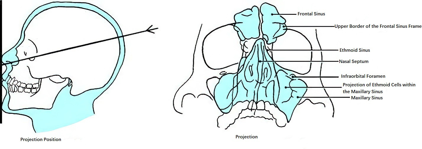
Figure 1 Nose-chin view
Occipital-Frontal View (Caldwell Position)
This is used primarily for examining the frontal and ethmoid sinuses, and also to visualize the maxillary sinuses, nasal cavity, and orbits.
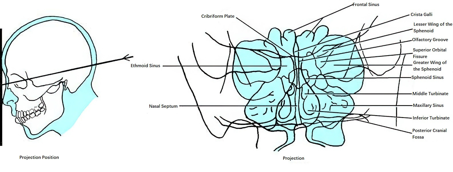
Figure 2 Occipital-frontal view
Additional Views as Needed
Lateral view (to observe various nasal sinuses, the sella turcica, and the nasopharynx), optic foramen view (to observe the ethmoid and sphenoid sinuses, and also the frontal sinus and orbital apex), and basal view (to observe the sphenoid sinus, posterior wall of the maxillary sinus, skull base, nasal cavity, and nasopharynx) are used.