The sinuses are located deeply and are relatively concealed. Routine anterior rhinoscopy, combined with positional drainage, maxillary sinus puncture, X-ray imaging, CT, and MRI, can help identify pathological changes either directly or indirectly.
Anterior Rhinoscopy
The goals of this examination include:
- Observing for any lesions in the nasal cavity that may obstruct drainage from the middle nasal meatus, such as high-grade deviations of the nasal septum or mucosal nodules.
- Examining nasal secretions for their color, characteristics, volume, and direction of drainage. In cases of anterior group sinusitis, purulent secretions are frequently observed draining from the middle nasal meatus, while in posterior group sinusitis, secretions often flow from the olfactory fissure toward the posterior nasal choanae, leading to postnasal drip symptoms.
- Noting whether polyps or neoplasms are present in the nasal meatus, and whether the mucosa of the nasal conchae exhibits swelling or polypoid changes. Enlargement of the uncinate process and ethmoid bulla are common signs of chronic sinusitis.
Positional Drainage Method
By identifying the source of purulent nasal secretions, the presence and specific location of sinusitis may be determined. Constriction of the nasal mucosa is achieved using 1% ephedrine to promote patency of the sinus ostia (e.g., those in the middle nasal meatus or the olfactory fissure). The patient remains in the required position for 15 minutes, after which an examination is conducted.
- For suspected maxillary sinus empyema, the head is tilted forward 90°, with the affected side oriented upward, to observe drainage in the posterior portion of the middle nasal meatus.
- For suspected frontal sinus empyema, the patient’s head is kept upright.
- For suspected anterior ethmoid sinus empyema, the head is slightly tilted backward.
- For suspected posterior ethmoid sinus empyema, the head is slightly leaned forward.
- For suspected sphenoid sinus empyema, the head is positioned downward with the forehead or nasal tip pressed against a flat plane.
Alternatively, the "head-low drainage method" may be used: The patient adopts a seated position with legs apart, leans the upper body forward, and allows the head to hang close to the knees. After approximately 10 minutes, the patient sits back up, and the nasal cavity is inspected for purulent drainage flowing into the nasal meatus.
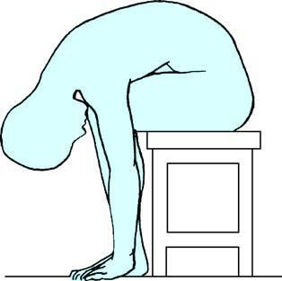
Figure 1 Head-low drainage method
Maxillary Sinus Puncture and Irrigation Method
This technique serves both diagnostic and therapeutic purposes. However, with the growing use of nasal endoscopy, this method is increasingly less utilized in clinical practice.
Imaging Techniques: X-Ray, CT, and MRI of the Sinuses
Details are provided in the relevant sections.