When sound waves cause vibrations in the basilar membrane of the cochlea, the outer hair cells, which exhibit frequency-specific characteristics, generate an active contraction response. These mechanical vibrations, initially triggered in the cochlea, propagate in reverse through the middle ear and the external auditory canal. This process likely serves to enhance the mechanical response of the basilar membrane to frequency-specific sound stimuli, allowing maximal vibration at corresponding locations and forming frequency-tuned traveling waves. The sound energy that originates from the cochlea and is transmitted to the external auditory canal via the ossicular chain and tympanic membrane is referred to as otoacoustic emission (OAE). OAE reflects the functional status of the outer hair cells in the cochlea. In addition to the attenuated stimulus sound, specialized high-sensitivity microphones can record sound energy delayed by several milliseconds within the external auditory canal.
Spontaneous otoacoustic emission (SOAE) refers to otoacoustic emissions recorded from the ear in the absence of sound stimulation. SOAEs are observed in approximately 40% of individuals with normal hearing. Evoked otoacoustic emission (EOAE) refers to otoacoustic emissions elicited by specific forms of sound stimulation in the test ear.
Evoked OAEs can be classified based on the type of stimulus sound:
- Transiently evoked otoacoustic emissions (TEOAEs) involve short-duration sound signals, such as single brief sounds or clicks, as the stimulus source.
- Stimulus-frequency otoacoustic emissions (SFOAEs) involve steady pure-tone signals used as the stimulus sound.
- Distortion product otoacoustic emissions (DPOAEs) are generated using two different pure-tone frequencies with a specific frequency ratio as a stimulus source. DPOAEs are the most commonly used type of OAE in clinical practice.
In individuals with normal hearing, the appearance rate of transiently evoked otoacoustic emissions is between 90% and 100%. Evoked otoacoustic emissions typically disappear when the pure-tone hearing threshold exceeds 30 dB HL. DPOAEs exhibit strong frequency specificity. Although they reflect outer hair cell function across the 1–8 kHz range, their sensitivity in the low-frequency region is limited, primarily reflecting outer hair cell function above 4 kHz. Therefore, combined analysis of TEOAEs and DPOAEs allows for a relatively accurate assessment of cochlear function.
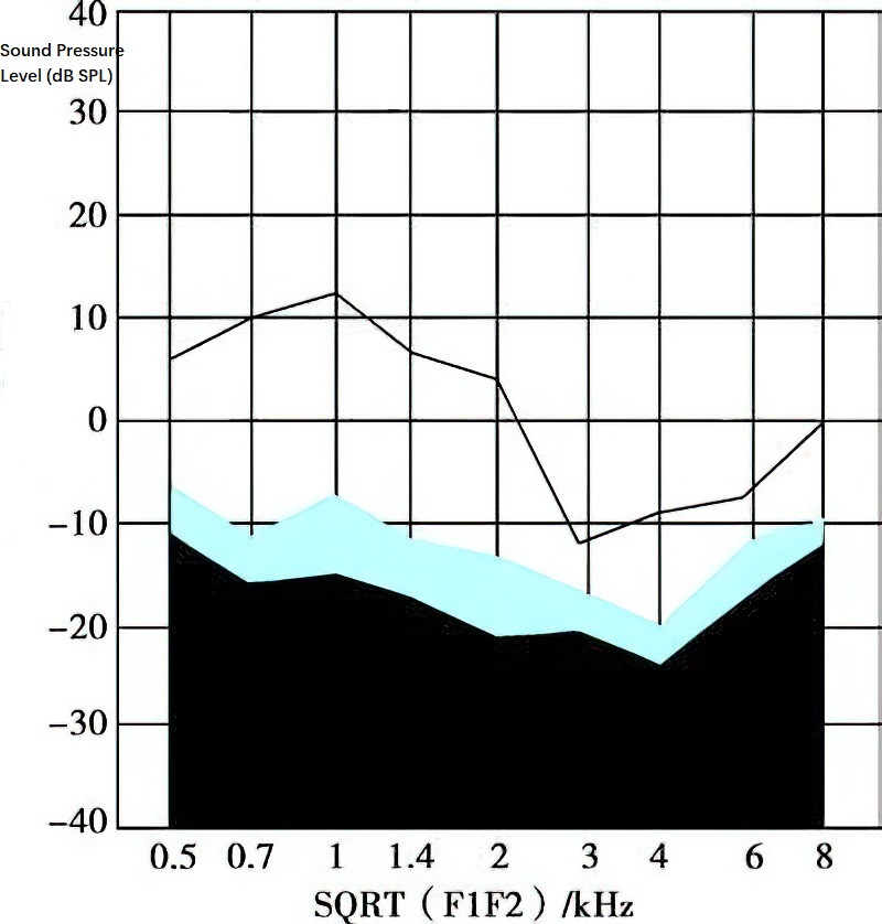
Figure 1 Normal distortion product otoacoustic emissions audiogram
The shaded area below represents background noise. Hearing response curves above the background noise baseline are considered normal.
An otoacoustic emissions graph is formed by connecting decibel values corresponding to different frequency sound energies. Decibel values greater than 10 dB above the background noise baseline are considered normal, whereas values below the baseline indicate no response. Because of its objectivity, simplicity, time efficiency, non-invasiveness, and sensitivity, OAE testing has become the preferred method for hearing screening in infants and young children. Infants who do not pass the OAE screening undergo further testing, such as auditory brainstem response (ABR). OAE testing is widely used in newborn hearing screening in maternity wards.
In cases where otoacoustic emissions are normal but auditory brainstem response is abnormal, the condition suggests auditory pathway disorders, such as auditory neuropathy or early-stage acoustic neuromas.