The basic examination of the Eustachian tube involves indirect observation of the nasopharynx using a mirror inserted through the oral cavity. Alternatively, a nasal endoscope or a thin fiberoptic endoscope may be inserted through the nasal cavity to observe the lumen of the Eustachian tube. A normal Eustachian tube is located on the lateral wall of the nasopharynx, with its pharyngeal opening surrounded by elevations and appearing light red in color.
In cases of nasopharyngeal inflammation, the elevations and the pharyngeal opening may appear red and swollen, with purulent nasal secretions from sinusitis potentially blocking the opening. Persistent secretory otitis media in children warrants examination for enlarged adenoids compressing the elevations and the pharyngeal opening. For individuals unable to cooperate with the examination, a lateral X-ray of the nasopharynx may serve as an alternative. In adults with unilateral secretory otitis media, the possibility of nasopharyngeal tumors compressing the Eustachian tube opening should be considered. Besides morphological assessment, the following methods can evaluate Eustachian tube function.
Eustachian Tube Insufflation
Airflow is directed actively or passively through the Eustachian tube into the tympanic cavity to assess Eustachian tube function in individuals without tympanic membrane perforation. This technique may also help relieve negative middle ear pressure or middle ear effusion. Its use is contraindicated in cases of acute upper respiratory infection, as well as in the presence of purulent discharge, ulcers, or neoplasms in the nasal cavity or nasopharynx.
Swallowing Test
The olive tips of a diagnostic stethoscope are placed at the ear canal entrances of the patient and the examiner. During the patient's swallowing motion, the examiner may hear a soft "hissing" sound transmitted through the stethoscope. Tympanic membrane movement corresponding to swallowing can also be observed via an otoscope. In cases of Eustachian tube dysfunction, the sound may not be heard, and tympanic membrane movement may be impaired. The Valsalva maneuver, also known as the "nose-pinching inflation method," directs more airflow into the middle ear through the Eustachian tube compared to the swallowing test.
Politzer Method
The Politzer method is used for individuals with poor Eustachian tube function or young children. The examiner places the olive tip of a Politzer bag into one of the patient's anterior nostrils and compresses the other nostril. When the patient swallows water, the soft palate elevates, closing the nasopharynx and momentarily opening the Eustachian tube. At this instant, the examiner compresses the rubber bag, directing airflow through the Eustachian tube into the tympanic cavity. The resulting sound of tympanic membrane vibration may be heard through a stethoscope, and tympanic membrane movement can be observed. This method can also be used therapeutically for Eustachian tube dysfunction.
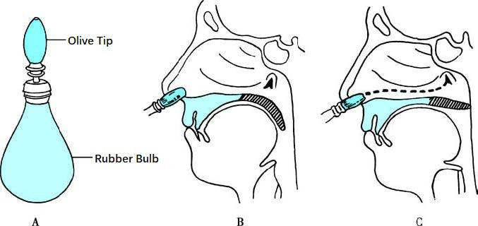
Figure 1 Politzer method
Catheter Inflation Method
After nasal mucosal vasoconstriction and anesthesia with 1% ephedrine and 1% tetracaine solutions, a Eustachian tube catheter is gently inserted along the nasal floor into the nasopharynx. The catheter is then rotated 90° towards the side being examined, withdrawn to the posterior nasal septum, and further rotated 45° upward so that its tip enters the pharyngeal opening of the Eustachian tube. Air is then insufflated into the catheter using a rubber bulb, with care to avoid excessive pressure that could rupture the tympanic membrane. A dual-balloon insufflation system may be employed to control pressure. This method is often used to treat Eustachian tube dysfunction and secretory otitis media.
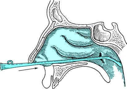
Figure 2 Step one of the Eustachian tube catheter insufflation method
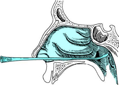
Figure 3 Step two of the Eustachian tube catheter insufflation method
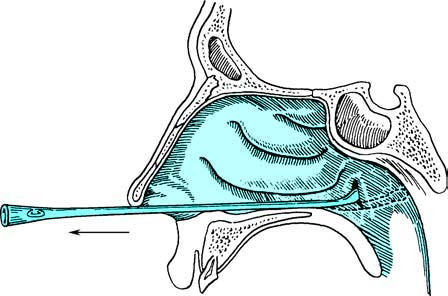
Figure 4 Step three of the Eustachian tube catheter insufflation method
Tympanic Instillation Method
This method is used to evaluate Eustachian tube function preoperatively in patients with tympanic membrane perforation. Specifically, a flavored solution such as ofloxacin ear drops or saccharin solution is instilled into the affected external auditory canal, and the patient is asked if they can taste the solution during swallowing. Alternatively, a sterile colored solution like methylene blue may be used, and the presence of the solution at the pharyngeal opening of the Eustachian tube is observed.
Eustachian Tube Radiography
Iodine-based contrast agents are instilled into the external auditory canal, allowing the agent to flow into the tympanic cavity through the tympanic membrane perforation. An X-ray examination is then performed to assess the anatomical structure of the Eustachian tube, the presence of narrowing or obstruction, and the natural drainage function.
Tympanometric Testing
Tympanometric pressure curves are obtained using an acoustic impedance instrument to evaluate Eustachian tube function. This method is non-invasive, objective, and quantitative. Details can be found in the section on tympanometric testing in this chapter.
Sonotubometry
Sonotubometry involves the use of a nasal probe to emit sound stimuli, with an external auditory canal probe detecting the transmitted sound. The data is analyzed by a computer to quantitatively assess the degree of Eustachian tube opening and functionality.
Tubomanometry
Tubomanometry (TMM) employs a pressure-detection device capable of sealing the external auditory canal, along with a system for generating and detecting pressure in the nasopharynx. The equipment generates adjustable pressure (30–50 mbar, where 1 mbar = 100 Pa) while simultaneously measuring changes in pressure within the nasopharynx and external auditory canal. During swallowing, the pressure changes are monitored and recorded to analyze the pressure dynamics on both sides of the Eustachian tube.