Anterior Chamber Angle and Gonioscope
Anterior Chamber Angle
The anterior chamber angle consists of three parts: the anterior wall, the posterior wall, and the recess between the two walls.
- Anterior Wall: The most anterior structure is Schwalbe’s line, which represents the termination of the corneal Descemet's membrane. It appears white, glossy, and slightly elevated. Posterior to Schwalbe's line is the trabecular meshwork, which typically has pigment deposition and serves as the main pathway for aqueous humor outflow. The Schlemm's canal lies external to the trabecular meshwork. The anterior wall ends at the scleral spur, which is white in color.
- Recess (Ciliary Body Band): This represents the anterior end of the ciliary body and appears black. It is also referred to as the ciliary body band.
- Posterior Wall: The posterior boundary is formed by the root of the iris.
Gonioscope
The gonioscope is an essential tool for directly observing the structures of the anterior chamber angle. It is frequently used in the diagnosis and management of conditions like glaucoma and ocular trauma. Gonioscopes function via the principles of light refraction (direct gonioscope) or reflection (indirect gonioscope). They are typically used in conjunction with surgical microscopes or slit-lamp biomicroscopes.
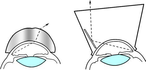
Figure 1 Direct gonioscope and indirect gonioscope
Clinical Description of Anterior Chamber Angle Width and Openness
Assessing the width and openness of the anterior chamber angle is critical for glaucoma diagnosis, classification, and treatment.
Anterior Chamber Angle Grading Systems
These grading systems are used to assess the width and either the openness or closure of the angle. The Scheie grading system is the most commonly employed clinically, although other systems, such as the Shaffer grading system and the Spaeth grading system, are also available.
Scheie Grading System
This system focuses on the most posterior structure of the angle recess that can be observed using gonioscopy. When the eye is in the primary position (static observation), an angle where all structures (Schwalbe's line, trabecular meshwork, scleral spur, and ciliary body band) are visible is categorized as a wide angle, while a narrower angle is classified as a narrow angle. Narrow angles are graded on a scale from Grade I to Grade IV as the angle progressively narrows:
- Grade I Narrow Angle: Slight narrowing.
- Grade II Narrow Angle: Further narrowing.
- Grade III Narrow Angle: Significant narrowing.
- Grade IV Narrow Angle: Almost completely closed.
In dynamic evaluation (e.g., altering eye position or applying slight pressure), the openness or closure of the angle can be determined. Observation of posterior trabecular structures indicates an open angle, while their absence suggests a closed angle.
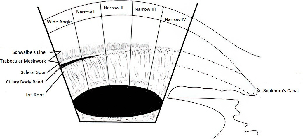
Figure 2 Scheie anterior chamber angle grading system