The perception of the three primary colors (red, green, and blue) by the human eye depends on the photopigments in the cone cells. Cone cells containing photopigments sensitive to red, green, and blue light are most responsive to wavelengths of 570 nm, 540 nm, and 440 nm, respectively. All colors can theoretically be created by combining red, green, and blue light in specific proportions, according to the trichromatic theory.
Individuals with normal proportions of the three photopigments are referred to as having trichromatic vision. Those with only two functioning types of photopigments are classified as having dichromatic vision, while individuals with only one functioning photopigment are classified as having monochromatic vision. Based on the trichromatic theory, individuals who can distinguish all three primary colors are considered to have normal color vision. Those who cannot distinguish any of the three primary colors have complete color blindness, while those unable to recognize one primary color are categorized as having dichromatic vision. Dichromatic vision results from the absence of a cone photopigment: individuals lacking red-sensitive photopigments have protanopia (red-blindness), while those lacking green-sensitive photopigments have deuteranopia (green-blindness). A reduced ability to perceive any given color is referred to as color deficiency, most commonly involving red (protanomaly) or green (deuteranomaly).
Color vision deficiency can be classified as congenital or acquired. Most congenital cases are X-linked recessive and predominantly affect men, with red-green deficiencies being the most common. Acquired color vision deficiencies may result from ocular or systemic conditions, such as optic nerve diseases, retinal disorders, drug toxicity, or media opacities such as corneal scars or cataracts.
Color vision testing is a routine part of pre-enrollment, employment, and military service physical examinations. It also serves as an auxiliary diagnostic tool for conditions like glaucoma and optic neuropathies. Furthermore, it is used to assess cone cell function before cataract surgery and to evaluate postoperative visual function. Color vision examination includes visual psychophysics-based testing (subjective methods) and visual electrophysiology-based testing (objective methods).
Pseudoisochromatic Plate Test
The pseudoisochromatic plate, commonly referred to as the color blindness test, is the simplest, fastest, and most widely used method of assessing color vision. Each plate contains figures or numbers formed by spots of different colors but similar brightness, or spots of the same color with different brightness. This test utilizes the confusion of certain color types to detect abnormalities. Individuals with normal color vision distinguish the figures by color, whereas colorblind individuals rely on brightness differences. Testing is conducted under natural light, with the plate positioned 0.5 meters from the examinee's eye. The examinee is required to identify the figure within 5 seconds.
Various pseudoisochromatic plates are available, catering to different testing needs. For example, Ishihara plates are commonly used for screening purposes. The AO-HRR test (American Optical Hardy-Rand-Rittler Color Vision Plates) provides semi-quantitative results, while SPP II (Standard Pseudoisochromatic Plates part 2) is utilized for assessing acquired color vision deficiencies.
Hue Arrangement Testing
This method involves arranging a set of color samples in order based on hue. The correctness of the sequence reflects the nature and degree of the individual's color vision deficiency. Common methods include the Farnsworth-Munsell (FM) 100-Hue Test and the Farnsworth D-15 Hue Test.
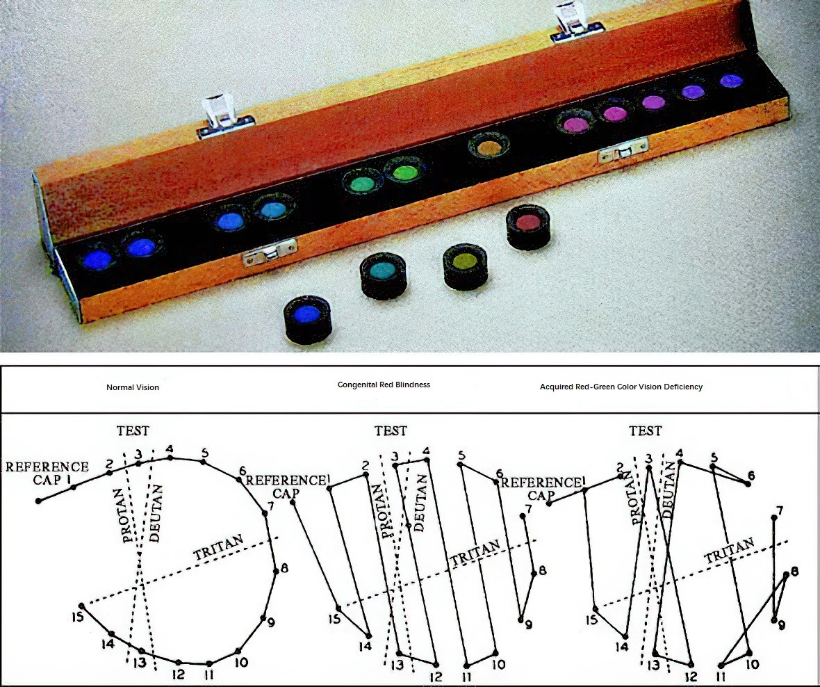
Figure 1 Farnsworth D-15 hue test
Anomaloscope Examination
The anomaloscope assesses color vision by utilizing the principle of mixing red and green light to create yellow light. Based on the examinee's ability to adjust the proportions of red and green light, the presence, nature, and severity of color vision deficiency can be determined.