Vessels
The blood vessels of the eye consist of the central retinal vascular system and the ciliary vascular system.
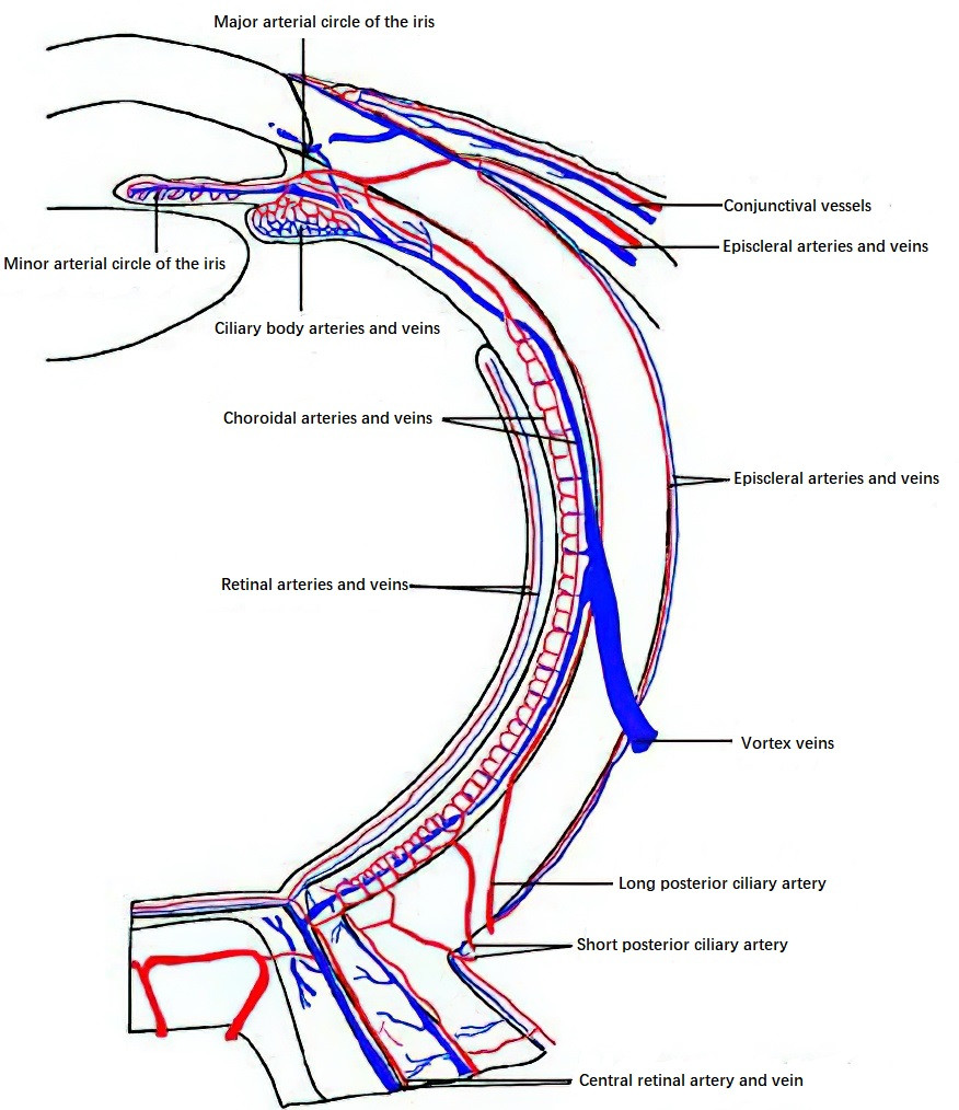
Figure 1 Diagram of ocular blood circulation
Central Retinal Artery (CRA)
The CRA is a branch of the intraorbital segment of the ophthalmic artery. It enters the optic nerve 9–12 mm posterior to the eyeball from the inferomedial or inferior side and emerges at the optic disc, where it divides into four branches: superior temporal, inferior temporal, superior nasal, and inferior nasal. These branches travel within the retinal nerve fiber layer and extend toward the peripheral retina. The central artery forms a capillary network through five levels of branching, which is further divided into superficial and deep layers. The superficial layer is found in the nerve fiber layer and the ganglion cell layer, while the deep layer is located in the inner nuclear layer. Avascular zones are present at the center of the macula. The CRA is an end artery and supplies the inner five layers of the retina. Around 30% of eyes also have cilioretinal arteries originating from the short posterior ciliary arteries, which supply the inner retinal layers. In only 15% of individuals, these arteries contribute to the blood supply of the macular portion.
Ciliary Vessels
The ciliary vessels are classified based on their location and course into short posterior ciliary arteries, long posterior ciliary arteries, and anterior ciliary arteries.
Short Posterior Ciliary Arteries
These are a group of branches from the ophthalmic artery, consisting of nasal and temporal trunks. They penetrate the sclera around the optic nerve and divide into about 20 branches, which enter the choroid and branch further down to the capillary level in a lobular distribution, providing nourishment to the choroid and the outer five retinal layers.
Long Posterior Ciliary Arteries
Two branches arise from the ophthalmic artery, located slightly distal to the nasal and temporal sides of the optic nerve. They obliquely traverse the sclera, entering the suprachoroid space, and extend anteriorly to the posterior portion of the ciliary body, where they start to branch. Some smaller branches return to the anterior choroid, while most branches extend to the anterior ciliary body and the posterior base of the iris. These branches connect with perforating branches of the anterior ciliary artery to form the major arterial circle, which gives rise to additional small branches that form the minor arterial circle near the pupillary margin. Small branches also extend to the ciliary muscle and processes, forming the vascular network of the ciliary body.
Anterior Ciliary Arteries
These derive from muscular branches of the ophthalmic artery. Near the tendon insertions of the extraocular muscles, these arteries travel within the scleral surface and stroma, giving rise to three types of branches. The episcleral branches travel anteriorly to the limbus, forming the limbal vascular arcade. The intrascleral branches penetrate the sclera and terminate around the Schlemm's canal. The perforating branches pass through the sclera to the ciliary body and contribute to the formation of the major arterial circle.
The blood supply to the optic disc is distinct in its characteristics. The optic disc's superficial nerve fiber layer is supplied by the capillaries of the CRA, while the pre-laminar and laminar regions are supplied by branches of the short posterior ciliary arteries, collectively known as the Zinn-Haller ring. This ring also communicates with the CRA.
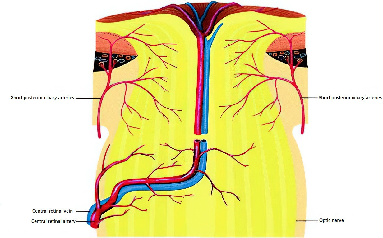
Figure 2 Schematic of the blood supply to the optic nerve
The venous drainage of the eye primarily involves:
Central Retinal Vein (CRV)
This vein accompanies the CRA and drains into the cavernous sinus either via the superior ophthalmic vein or directly.
Vortex Veins
These veins are located posterior to the equator of the eyeball and collect venous blood from the choroid as well as parts of the iris and ciliary body. There are 4–7 vortex veins, 1–2 per quadrant of the eye. They exit the sclera obliquely through the spaces between the rectus muscles, located 14–25 mm from the corneal limbus. The vortex veins drain into the cavernous sinus via the superior and inferior ophthalmic veins.
Anterior Ciliary Veins
These veins collect blood from the iris and ciliary body. Venous blood from the upper half flows into the superior ophthalmic vein, while blood from the lower half flows into the inferior ophthalmic vein. Most of the blood is eventually drained into the cavernous sinus via the superior orbital fissure, while a portion drains into the facial vein and the pterygoid venous plexus through the inferior orbital fissure, finally entering the external jugular vein.
Nerves
The nerve supply to the eye is extensive, involving six pairs of cranial nerves. These include the optic nerve (cranial nerve II), the oculomotor nerve (cranial nerve III), the trochlear nerve (cranial nerve IV), the trigeminal nerve (cranial nerve V), the abducens nerve (cranial nerve VI), and the facial nerve (cranial nerve VII).
The optic nerve (cranial nerve II) is responsible for visual signal transmission. The oculomotor nerve (cranial nerve III) innervates all intraocular muscles, the levator palpebrae superioris, and all extraocular muscles except the lateral rectus and superior oblique muscles. The trochlear nerve (cranial nerve IV) innervates the superior oblique muscle, while the abducens nerve (cranial nerve VI) innervates the lateral rectus muscle. The trigeminal nerve (cranial nerve V) is responsible for sensations in the eye, and the facial nerve (cranial nerve VII) innervates the orbicularis oculi muscle. Unique neural structures are also formed within the orbit through the interaction of cranial nerves III and V with the autonomic nervous system.
Ciliary Ganglion
The ciliary ganglion is located lateral to the optic nerve, approximately 10 mm anterior to the common tendinous ring. It receives preganglionic fibers from three roots:
- The long root (sensory root), which originates from the nasociliary nerve.
- The short root (motor root), which originates from the oculomotor nerve (cranial nerve III) and contains parasympathetic fibers.
- The sympathetic root, which arises from the internal carotid plexus and controls the vasoconstriction and dilation of ocular blood vessels.
Postganglionic fibers from the ciliary ganglion form the short ciliary nerves. Retrobulbar anesthesia, performed during intraocular surgeries, blocks this ganglion.
Nasociliary Nerve
The nasociliary nerve is a branch of the ophthalmic division of the trigeminal nerve (cranial nerve V) and is responsible for sensory innervation of the eye. Within the orbit, it gives rise to several branches, including the sensory root of the ciliary ganglion, long ciliary nerves, posterior ethmoidal nerve, and infratrochlear nerve.
The long ciliary nerves emerge as two branches posterior to the eyeball, passing along both sides of the optic nerve and piercing the sclera. These nerves contain sympathetic fibers that join them and travel within the suprachoroidal space. They carry corneal sensory input and sympathetic fibers to the ciliary muscle and the dilator pupillae muscle.
The short ciliary nerves are mixed nerves, numbering 6–10 branches, which pierce the sclera around the optic nerve in the posterior pole of the eyeball. They also travel within the suprachoroidal space and extend to the ciliary body, where they form a plexus. From this plexus, branches emerge to innervate the iris, ciliary body, cornea, and sclera. Parasympathetic fibers are conveyed to the sphincter pupillae and ciliary muscles, while sympathetic fibers extend to the intraocular blood vessels, regulating vascular dilation and constriction.