The visual pathway refers to the conduction route through which visual information is transmitted from the photoreceptors of the retina to the visual cortex in the occipital lobe of the brain. Clinically, it generally refers to the neural conduction pathway that starts from the optic nerve, passes through the optic chiasm, optic tract, lateral geniculate body, optic radiation, and finally reaches the visual cortex in the occipital lobe.
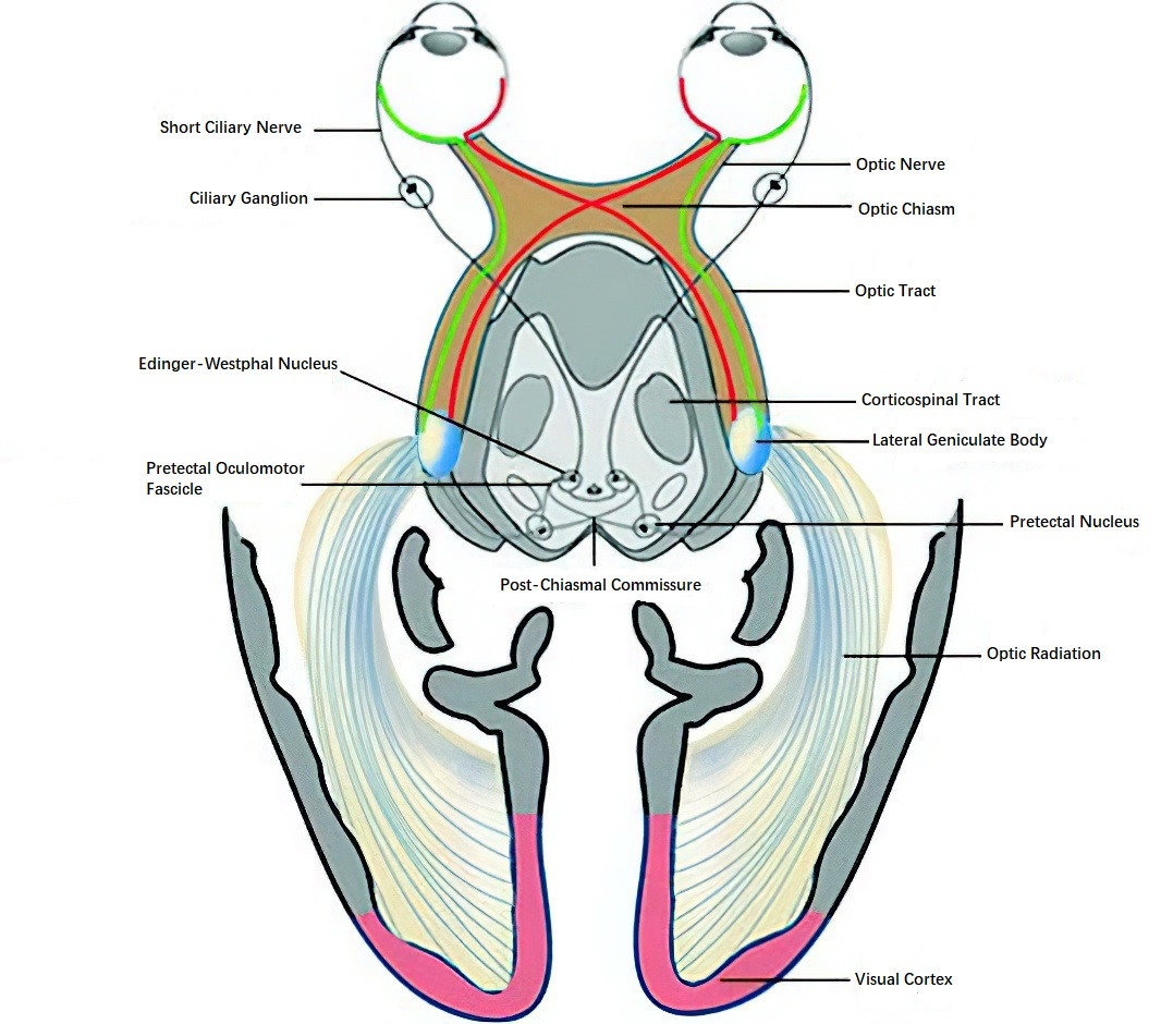
Figure 1 Diagram of the visual pathway
Optic Nerve
The optic nerve is part of the central nervous system. The segment of the nerve extending from the optic disc to the anterior portion of the optic chiasm is referred to as the optic nerve, with an average total length of about 40 mm. It is divided into four segments based on location: intraocular, intraorbital, intracanalicular, and intracranial segments.
Intraocular Segment
This segment begins at the optic disc, where the axons of 1–1.2 million ganglion cells bundle together to form the nerve fibers and traverse the scleral lamina cribrosa to exit the eyeball. It is about 1 mm in length and consists of four parts: the nerve fiber layer, prelaminar layer, laminar layer, and retrolaminar area. Clinically, the nerve fiber layer (reddish-orange in appearance) and the prelaminar central part (optic cup) can be observed on fundus examination. Occasionally, small gray dot-like pores at the base of the optic cup can also be seen, which correspond to the lamina cribrosa. The prelaminar nerve fibers lack myelin sheaths (1.5 mm in diameter), whereas myelination begins after they pass through the lamina cribrosa, increasing the diameter to 3.0 mm. The blood supply to the intraocular segment of the optic nerve is derived from branches of the retinal artery and posterior short ciliary arteries.
Intraorbital Segment
This segment is about 25 mm long and is located within the muscle cone. The optic nerve is enveloped by the optic nerve sheath, which is a continuation of the three layers of the meninges. The subarachnoid space within the sheath communicates with the intracranial subarachnoid space and is filled with cerebrospinal fluid. At a distance of 10–15 mm from the eyeball, fibers from the papillomacular bundle gradually shift to the axial portion of the optic nerve, while fibers from other parts of the retina remain in their corresponding locations within the optic nerve. The blood supply to the intraorbital segment of the optic nerve is primarily provided by branches of the ophthalmic artery and the central retinal artery.
Intracanalicular Segment
This segment refers to the portion of the optic nerve that passes through the optic canal of the skull, measuring 4–9 mm in length. The optic nerve sheath is tightly connected to the periosteum in this region, which helps fix the optic nerve in place. The blood supply is provided by the accompanying ophthalmic artery, and the arrangement of the nerve fibers remains unchanged in this segment.
Intracranial Segment
This segment extends from the optic canal to the anterior portion of the optic chiasm and measures about 10 mm in length, with a diameter of 4–7 mm. Its blood supply is provided by the internal carotid artery and ophthalmic artery.
Optic Chiasm
The optic chiasm is the crossing point of the optic nerves from both eyes. It has a rectangular shape, measuring about 12 mm in width, 8 mm in anteroposterior diameter, and 4 mm in thickness. The nerve fibers within the optic chiasm divide into two groups: nasal fibers from the retina cross to the opposite side, while temporal fibers do not cross. Yellow spot (macular) fibers comprise 80–90% of the central region of the optic nerve and optic chiasm, and they are also divided into crossing and non-crossing fibers.
The anatomical relationship of the optic chiasm with surrounding structures includes the anterior cerebral artery and anterior communicating artery superiorly and anteriorly, the internal carotid artery on both sides, the pituitary gland inferiorly, and the third ventricle posteriorly and superiorly. Pathological changes in these regions can affect the optic chiasm, leading to characteristic visual field defects.
Optic Tract
The optic tract refers to the segment of the optic nerve fibers that are rearranged after passing through the optic chiasm. It splits into two bundles that wrap around the cerebral peduncles to reach the lateral geniculate body. Nerve fibers from the inferior half of the retina (both crossed and uncrossed) occupy the lateral portion of the optic tract, while fibers from the superior half of the retina (both crossed and uncrossed) occupy the medial portion. Initially, macular fibers are located centrally but later shift to the dorsolateral portion of the optic tract.
Lateral Geniculate Body
The lateral geniculate body, located on the lateral side of the cerebral peduncle, has an oval shape. Approximately 70% of the nerve fibers from the ganglion cells of the retina form synapses with neurons in the lateral geniculate body, transitioning to the fourth-order neurons in the visual pathway before entering the optic radiation. The gray and white matter within the lateral geniculate body are arranged in alternating layers. The crossed fibers from the contralateral retina terminate in layers 1, 4, and 6, while the uncrossed fibers from the ipsilateral retina terminate in layers 2, 3, and 5.
Optic Radiation
The optic radiation consists of the neural fibers connecting the lateral geniculate body to the occipital cortical area. After transitioning to the next set of neurons, these nerve fibers pass through the internal capsule and the posterior-inferior portion of the lentiform nucleus, spreading out in a fan-like shape in three bundles (dorsal, lateral, and ventral) around the temporal horn of the lateral ventricle, forming Meyer’s loop, and eventually reaching the occipital lobe.
Visual Cortex
The visual cortex is located in the occipital lobe and corresponds to Brodmann areas 17, 18, and 19, including the lips of the calcarine fissure and the striate area of the occipital lobe. It is the thinnest region of the cerebral cortex. Each side of the visual cortex is associated with one-half of the retina from both eyes: for instance, the left visual cortex is linked to the temporal half of the left eye and the nasal half of the right eye's retina. Nerve fibers from the upper retina terminate along the upper lip of the calcarine fissure, while fibers from the lower retina terminate along the lower lip. Macular fibers terminate at the posterior pole of the striate area in the occipital lobe. Crossed fibers are located in the deep internal granular layers, while uncrossed fibers are located in the superficial internal granular layers.
Since the arrangement of visual nerve fibers varies along different segments of the visual pathway, lesions or damage in specific parts of the nervous system result in corresponding visual field defects. Identifying these characteristic visual field changes is crucial for the accurate localization and diagnosis of central nervous system disorders.