The appendix is located in the right iliac fossa, appearing worm-like in shape. Its length varies between 2 cm and 20 cm, with an average of 6–8 cm, and a diameter of 0.5–0.7 cm. It originates at the distal end of the cecum and is attached at the convergence point of the three taeniae coli of the colon. This anatomical feature enables the root of the appendix to be located during surgery by tracing the taeniae coli to the cecum. The surface projection of the appendix is approximately at the junction of the medial two-thirds and the lateral one-third along the line connecting the umbilicus and the right anterior superior iliac spine, known as McBurney's point. McBurney's point serves as a landmark for an incision during appendectomy procedures. Most appendices are intraperitoneal organs, though their positions can vary significantly.
Due to the fixed relationship between the appendix root and the cecum, the position of the appendix changes depending on the location of the cecum. It is typically found in the lower right quadrant of the abdomen but can extend to lie beneath the liver, descend into the pelvic cavity, or even traverse the midline to the left side. The anatomical position of the appendix can vary like the hands of a clock around the root, which serves as the center of this 360-degree reference. This variability explains differences in clinical symptoms and tenderness points in patients. The tip of the appendix has six common positional types:
- Ileocecal anterior position: Located between the "12 o'clock" and "3 o'clock" positions, with the tip pointing upward to the left.
- Pelvic position: Corresponding to the "3 o'clock" to "6 o'clock" positions, with the tip directed toward the pelvis.
- Retrocecal position: Found between the "9 o'clock" and "12 o'clock" positions, located behind the cecum and anterior to the iliacus muscle, with its tip pointing upward and lying retroperitoneal. Appendicitis in this position often presents with mild clinical signs and can be misdiagnosed. Exposing and removing the appendix in this position during surgery can be challenging.
- Infra-cecal position: Corresponding to the "6 o'clock" to "9 o'clock" positions, with its tip pointing downward to the right.
- Laterocecal position: Located between the "9 o'clock" and "10 o'clock" positions, situated intraperitoneally on the lateral side of the cecum.
- Post-ileal position: Found between the "12 o’clock" and "3 o’clock" positions, but situated posterior to the ileum.
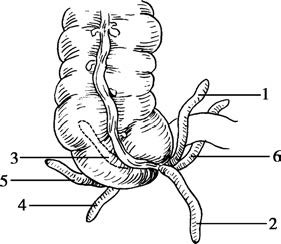
Figure 1 Anatomical positions of the appendix
1, Ileocecal anterior position
2, Pelvic position
3, Retrocecal position
4, Infra-cecal position
5, Laterocecal position
6, Post-ileal position
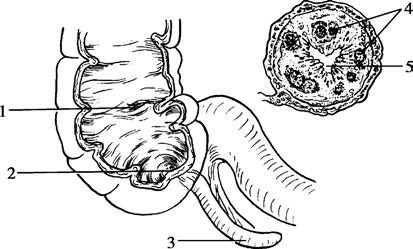
Figure 2 Anatomy of the appendix
1, Ileocecal valve
2, Appendiceal orifice
3, Appendix
4, Lymphoid tissue
5, Appendiceal lumen
The appendix is a tubular organ with a blind-ended distal tip and a proximal opening into the cecum, located 2–3 cm below the ileocecal valve. Its mesoappendix is triangular or fan-shaped and contains blood vessels, lymphatic vessels, and nerves. The mesoappendix is shorter than the appendix itself, which leads to the appendix appearing coiled. The vessels in the mesoappendix primarily consist of the appendicular artery and vein, which pass along the free border of the mesoappendix posterior to the terminal ileum. The appendicular artery is a branch of the ileocolic artery and is classified as an end artery with no collateral circulation, making the appendix susceptible to ischemia and subsequent necrosis in the event of vascular obstruction. The appendicular vein runs alongside the artery and eventually drains into the portal vein. Inflammation in the appendix can lead to bacteremia, which may cause pylephlebitis or bacterial liver abscesses.
The appendix's lymphatic vessels accompany the blood vessels in the mesoappendix, eventually draining into the lymph nodes near the right colic artery, the anterior duodenal area, and the mesocolic nodes around the superior mesenteric artery. The appendix is innervated by sympathetic fibers that travel through the celiac plexus and lesser splanchnic nerves. Pain from acute appendicitis is referred to the periumbilical region due to its visceral nerve supply originating from the T10–T11 spinal segments.
The tissue structure of the appendix wall resembles that of the colon. The epithelial cells of the appendiceal mucosa are capable of secreting a small amount of mucus. The appendix is also a lymphoid organ and plays a role in the generation and maturation of B lymphocytes, contributing to immune functions. The appendix contains abundant lymphatic tissue and is thought to generate lymphocytes and antibodies, along with Peyer's patches in the terminal ileum, to help protect against infections such as viruses. Lymphatic tissue in the appendix begins to develop after birth and reaches its peak activity between the ages of 12 and 20, containing over 200 lymphoid follicles. This tissue gradually diminishes after the age of 30 and disappears entirely by the age of 60.