The adult female breast is a pair of hemispherical secondary sexual characteristics located on the superficial surface of the pectoralis major muscle, roughly spanning the second to sixth ribs. The upper outer quadrant extends into the axilla as the axillary tail of the breast. The nipple is situated centrally on the breast and is surrounded by a pigmented area called the areola.
The mammary gland is composed of 15 to 20 lobes, which are further divided into smaller lobules. Each lobule consists of terminal ducts and acini. The small ducts within the lobules converge to form lactiferous ducts for each lobe, which open onto the nipple. Both the lobes and the ducts are radially arranged around the nipple. The proximal third of the lactiferous ducts near the opening expands slightly, forming the lactiferous sinus. Between the lobes, lobules, and acini, connective tissue septa are present. Additionally, vertical fibrous bands, known as Cooper's ligaments, extend from the skin to the deep layer of the superficial fascia, providing structural support and anchoring the breast.
The mammary gland is a target organ for various endocrine hormones, and its physiological activity is influenced by hormones secreted by the pituitary gland, adrenal cortex, and ovaries. The breast undergoes different changes at various stages of life.
The blood supply to the breast is primarily provided by branches of the lateral thoracic vessels and internal thoracic vessels.
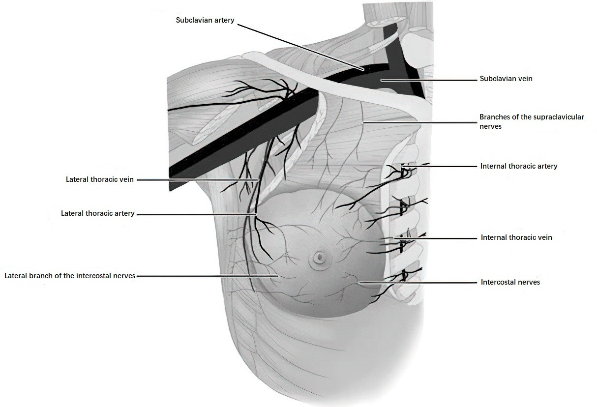
Figure 1 Blood supply and innervation of the breast
Innervation of the breast is mainly derived from branches of the supraclavicular nerves and the second to sixth intercostal nerves.
The lymphatic network of the breast is extensive, with lymphatic drainage occurring via four main pathways:
- The majority of lymphatic fluid from the breast drains into the axillary lymph nodes. Lymph from the upper portion of the breast can directly reach the subclavian lymph nodes.
- Lymph from the medial portion of the breast drains into the parasternal lymph nodes via intercostal lymphatic vessels.
- Subcutaneous communicating lymphatic vessels exist between the two breasts.
- The deep lymphatic network of the breast can drain along the rectus abdominis sheath and the falciform ligament of the liver, eventually linking to the liver.
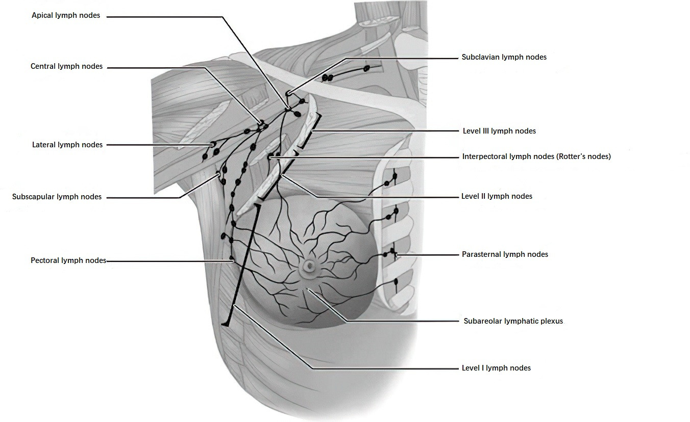
Figure 2 Axillary lymph node groups
Axillary lymph nodes are commonly classified into three levels based on their relationship to the pectoralis minor muscle:
- Level I: Lymph nodes lateral to the pectoralis minor muscle.
- Level II: Lymph nodes located posterior to the pectoralis minor muscle, including lymph nodes between the pectoralis major and minor muscles (Rotter's nodes).
- Level III: Lymph nodes medial to the pectoralis minor muscle, including subclavian lymph nodes.