History of Endoscopic Technology Development
The term "endoscopy" originates from Greek, referring to the method of observing the luminal structures within the human body. From its initial concept, endoscopy has undergone over two centuries of evolution and transformation, progressing from early rigid, semi-flexible, and fiberoptic endoscopes to today's electronic endoscopes. This development has established a comprehensive system that has revolutionized the diagnosis and treatment of diseases affecting the gastrointestinal, respiratory, and genitourinary systems.
Basic Principles and Types of Endoscopes
With technological advancements, most modern endoscopes use the principle of electronic endoscopy. By utilizing a charge-coupled device (CCD) at the tip of the endoscope, light signals are converted into electrical signals, which are then processed by a video system and displayed as images on a monitor.
Endoscopes are classified into rigid and flexible types:
Rigid Endoscopes
This category includes instruments such as cystoscopes, anoscopes, rectoscopes, thoracoscopes, arthroscopes, and ventriculoscopes. These endoscopes have a fixed structure and are non-bendable. Although they cannot freely adjust the viewing angle, they offer advantages such as ease of operation and greater durability. Rigid endoscopes can access the body cavity via natural orifices or through percutaneous trocar-assisted pathways.
Flexible Endoscopes
Instruments in this category include gastroscopes, colonoscopes, enteroscopes, duodenoscopes, cholangiopancreatoscopes, bronchoscopes, and nasopharyngoscopes. The shafts and tips of these endoscopes are bendable, allowing operators to perform procedures under direct visualization.
Common Diagnostic Techniques and Therapeutic Instruments in Endoscopy
Common Endoscopic Diagnostic Techniques
Chromoendoscopy and Magnification Endoscopy
Chromoendoscopy involves the application of special dyes to stain the gastrointestinal mucosa, enhancing disease detection rates. Magnification endoscopy can enlarge the observation field by 60 to 170 times. Combining chromoendoscopy with magnification endoscopy enables precise delineation of lesion characteristics and boundaries, thereby improving diagnostic accuracy. Staining mucosal lesions facilitates localization during procedures such as laparoscopy.
Electronic Chromoendoscopy
Narrow-band imaging (NBI) is commonly employed in electronic chromoendoscopy. It filters the broad-spectrum red, green, and blue light emitted by the endoscope’s light source into narrow-band wavelengths to illuminate the mucosa. This technique enhances the contrast and clarity of mucosal epithelium and submucosal blood vessels. It allows for detailed observation of early mucosal lesions, inflammatory changes, and the vascular architecture of tumor surfaces.
Endoscopic Imaging Techniques
Techniques such as endoscopic retrograde cholangiopancreatography (ERCP) and retrograde ureteropyelography increase diagnostic accuracy.
Pathological Biopsy
Tissue samples can be obtained using biopsy forceps during endoscopy for histopathological analysis. This plays a foundational role in planning subsequent treatments.
Common Therapeutic Energy Devices Used in Endoscopy
High-Frequency Electrosurgical Knife
This device is used for cutting tissue in electrosurgical procedures. Upon contact with biological tissue, high-frequency and high-voltage electric currents generated at the electrode tip instantaneously heat the tissue, enabling tissue dissection and coagulation for cutting and hemostasis.
Laser
Lasers are characterized by high brightness, monochromaticity, and strong directionality. They are utilized for cutting, coagulating, achieving hemostasis, and vaporizing tissue. Normal tissues and pathological lesions, such as tumors, emit different wavelengths of fluorescence upon laser excitation, aiding in the early localization of tumors.
Microwave
Microwaves, a type of electromagnetic wave with a frequency range of 300–300,000 MHz, cause polar molecules in biological tissue to rotate rapidly in response to alternating electric fields. The resulting thermal effects are used to treat certain mucosal diseases.
Radiofrequency (RF)
RF is a high-frequency alternating electromagnetic wave. RF currents greater than 10 kHz cause ions within biological tissue to vibrate, generating localized temperatures of 90–100°C around the electrode. This thermal conduction causes tissue ablation in the target area.
Argon-Helium Knife
This device rapidly brings the temperature of target tissues down to below -140°C within 10–20 seconds, followed by rapid warming to 30–35°C. This extreme temperature cycling results in the destruction of pathological tissues.
Applications of Endoscopic Technology in Surgery
Applications in Gastrointestinal Surgery
Gastroscopy
Gastroscopy enables procedures using high-frequency electrosurgical knives for treating lesions, such as through endoscopic mucosal resection (EMR) or endoscopic submucosal dissection (ESD). The advancement of techniques like endoscopic balloon dilation and stent placement provides minimally invasive treatment options for conditions such as esophageal strictures caused by tumors or anastomotic strictures post-surgery. Gastroscopy can also effectively treat upper gastrointestinal bleeding through techniques such as electrocoagulation, ligation, or local injection.
Colonoscopy
Similar to gastroscopy, colonoscopy is used to treat colorectal polyps and early-stage cancers. Intestinal obstructions caused by tumors can also be temporarily relieved using catheters or stents, creating favorable conditions for subsequent surgeries.
Enteroscopy
Enteroscopy, which includes double-balloon and single-balloon types, is utilized for diagnosing gastrointestinal bleeding, radiation-induced small bowel injury, and small bowel lesions not identified by capsule endoscopy.
Duodenoscopy
Duodenoscopy enables procedures such as ERCP, the management of bile duct stones, and endoscopic sphincterotomy (EST).
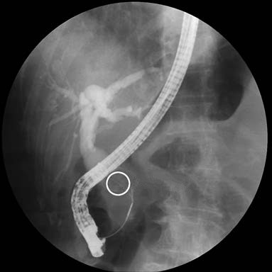
Figure 1 Endoscopic retrograde cholangiopancreatography (ERCP) demonstrating common bile duct stones
Cholangiopancreatoscopy
This endoscopic approach facilitates bile duct exploration, biopsy, and hemostasis. It is also used for retrieving bile duct stones and, in combination with balloon-assisted techniques, for bile duct dilation.
Ultrasonic Endoscopy
Ultrasonic endoscopy combines an endoscope with an ultrasound probe, allowing for ultrasound scanning of the walls of the gastrointestinal tract and surrounding organs. It is commonly used for tumor staging in the digestive tract, localization of submucosal tumors, and diagnosis of biliary and pancreatic diseases.
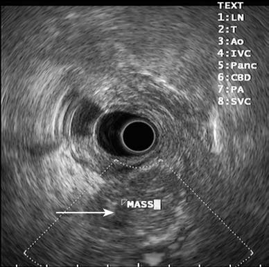
Figure 2 Pancreatic mass under endoscopic ultrasound
Endoscopic ultrasound reveals a hypoechoic mass with irregular borders.
Capsule Endoscopy
Capsule endoscopy involves swallowing a capsule equipped with an internal camera and wireless transmission device. The capsule travels through the gastrointestinal tract, capturing images that are viewed on an external imaging workstation for diagnostic purposes.
Confocal Laser Endomicroscopy
A small confocal laser microscope is attached to the tip of a standard endoscope, allowing real-time microscopic observation of living tissue at a histological level. This technology enables the simultaneous performance of endoscopic examination and tissue histology evaluation by visualizing fine structural details.
Applications in Urological Surgery
Endoscopic technology is widely used in urology, with most urinary system stones treatable through endoscopy. Percutaneous nephroscopy, ureteroscopy, cystoscopy, or laparoscopy can be combined with methods such as pneumatic lithotripsy, electrohydraulic lithotripsy, ultrasound, or laser therapy for stone treatment. Transurethral resection of the prostate (TURP) has become the standard approach for treating benign prostatic hyperplasia. For bladder cancer, different endoscopic treatments can be chosen based on its stage. For instance, superficial bladder cancer can be treated with transurethral resection of bladder tumors.
Applications in Thoracic Surgery
Bronchoscopy is primarily used in thoracic surgery for the diagnosis and removal of bronchial lesions, hemostasis, or balloon dilation in cases of bronchial stenosis.
Applications in Orthopedic Surgery
Knee arthroscopy is used to examine the internal structures of the joint, such as the synovium, cartilage, meniscus, and ligaments, and to treat joint disorders. Spinal endoscopy is another application, enabling minimally invasive spinal surgeries with advantages such as reduced tissue damage, minimal bleeding, less post-operative pain, shorter hospital stays, and less disruption to spinal stability.
Applications in Neurosurgery
Endoscopic technology in neurosurgery is commonly used for treating conditions such as hydrocephalus, intracranial cysts, intracranial hematomas, intraventricular and periventricular tumors, pituitary adenomas, and craniopharyngiomas.
Overview of Natural Orifice Translumenal Endoscopic Surgery (NOTES)
Natural Orifice Translumenal Endoscopic Surgery (NOTES) refers to a surgical approach where an endoscope is introduced through natural orifices (e.g., the mouth, anus, or vagina) to penetrate the mucosa at appropriate sites and access the thoracic, abdominal, or cervical cavities. Resected specimens are then extracted through the same natural orifice.
Attempts to perform NOTES began in the late 1980s with procedures like gallbladder removal and appendix removal. By the early 21st century, reports emerged of procedures involving vaginal oophorectomy, vaginal appendectomy, and transoral thyroidectomy. However, the broader implementation of NOTES remains contingent upon further advancements in endoscopic technology, specialized surgical instruments, and optimization of protocols for ensuring clear visualization of the surgical field. Currently, most NOTES procedures remain in the experimental stage.