Fibrous dysplasia of bone, also known as fibrous bone abnormal proliferation disease, is a developmental bone disorder. In this condition, bone development halts at the immature woven bone stage and does not progress to form normal trabecular bone. It can affect a single bone (monostotic) or multiple bones (polyostotic). It commonly occurs in adolescents and middle-aged individuals, most frequently between the ages of 10 and 25 during active skeletal growth. The marrow cavity of the affected bone contains fibrous tissue. The lesion consists of dense fibrous tissue with a disordered and non-oriented arrangement. Metaplastic bone, appearing as fibrous or woven bone, is present within the fibrous connective tissue. In some cases, the lesion may also show myxoid degeneration, multinucleated giant cells, and cartilage islands.
Clinical Features
The progression of the lesion is typically slow and often asymptomatic. It is frequently detected incidentally during X-ray imaging. Pathological fractures are a common complication. Radiographically, the affected bone appears enlarged and thickened, with thinning of the cortical bone. A classic feature is the "ground-glass" appearance with well-defined margins. Lesions in the proximal femur can lead to bending of the femoral neck, creating a deformity resembling a "shepherd's crook."
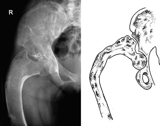
Figure 1 "Shepherd’s crook" deformity in fibrous dysplasia of the proximal femur
Treatment
Curettage with bone grafting can be performed. For certain long bones, such as the fibula or ribs, segmental resection may be considered. For cases with deformities, corrective osteotomy may be performed.