A bone cyst is an intramedullary lesion that is typically single-chambered, cystic, and localized, resembling a tumor-like condition. The cyst cavity contains serous or serosanguinous fluid. It is most commonly seen in children and adolescents, with a predilection for the metaphyses of long tubular bones. The most frequent sites, in order, are the proximal humerus, proximal femur, proximal tibia, and distal radius.
Clinical Features
Most cases are asymptomatic, though some patients may experience mild local pain or swelling. The majority seek medical attention only after a pathological fracture occurs. On X-ray, the lesion appears as a well-defined, round or oval lytic lesion at the metaphysis with cortical thinning due to expansion. The lesion is typically unilocular or multilocular and is often adjacent to the growth plate but does not cross it.
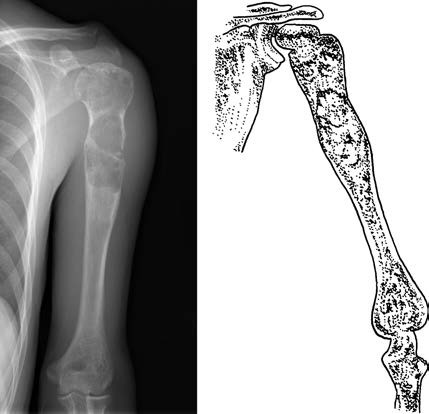
Figure 1 Bone cyst in the proximal humerus with pathological fracture
Treatment
The standard treatment for a simple bone cyst involves curettage of the lesion followed by filling the defect with autograft or allograft bone. Some bone cysts can heal spontaneously after a fracture. For younger children (under 14 years of age) or lesions located near the growth plate, surgical intervention is approached cautiously due to the risk of growth plate damage and a higher likelihood of local recurrence after surgery. Injecting methylprednisolone into the cyst cavity has shown some efficacy in restoring normal bone structure.