Osteosarcoma is the most common malignant bone tumor, characterized by the production of osteoid matrix by tumor cells. It includes various subtypes as well as secondary osteosarcoma. This tumor primarily affects adolescents and is most frequently located in the metaphyseal region of the distal femur, proximal tibia, and proximal humerus. It typically forms a spindle-shaped mass, involving the periosteum, cortical bone, and medullary cavity. On gross examination, the lesion appears fish-flesh-like, with a tan-reddish or greyish-white surface.
Clinical Features
The main symptom is localized pain, which is usually persistent, gradually worsens, and intensifies at night. In some cases, there is an associated palpable mass, restricted joint motion near the lesion, and localized warmth with dilated superficial veins overlying the tumor. Systemic manifestations of cachexia may also be present. Lytic osteosarcoma can erode the cortical bone, leading to pathological fractures. Radionuclide bone scanning assists in determining the tumor size and identifying metastatic lesions. Laboratory tests are useful for evaluating the status of the disease.
Radiological Features
X-ray imaging demonstrates variable morphologies, with bone destruction in a sclerotic, lytic, or mixed pattern involving both the cortical bone and medullary cavity. Periosteal reactions are prominent and aggressive, often presenting as Codman’s triangle or a “sunburst” appearance. MRI provides detailed delineation of tumor margins and the extent of invasive involvement.
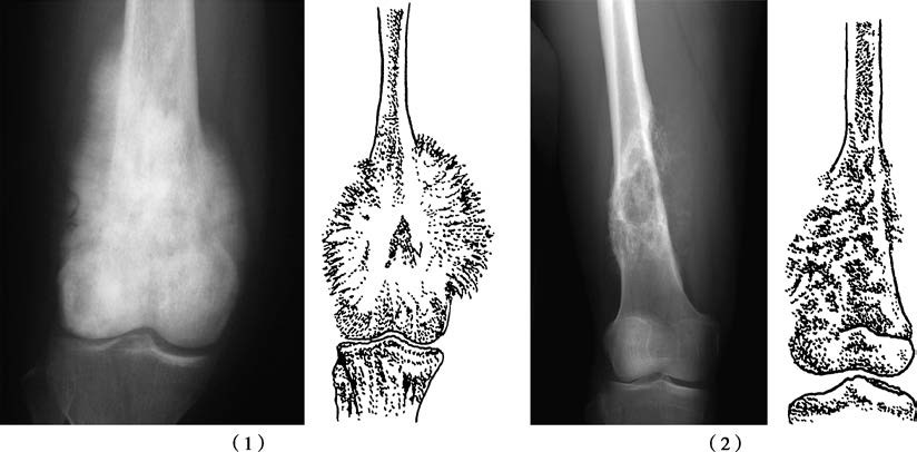
Figure 1 Osteosarcoma of the femur
(1) Distal femoral osteosarcoma showing a "sunburst" appearance
(2) Tumor in the distal femur, demonstrating osteoid production with bone destruction
Treatment
For cases classified as G2T1–2M0, multimodal therapy is employed. High-dose chemotherapy is administered preoperatively, followed by limb-sparing surgery with radical tumor resection and prosthesis implantation, or amputation, depending on the tumor’s extent of invasion. Postoperative high-dose chemotherapy is continued. Osteosarcoma has a high rate of pulmonary metastasis. For cases classified as G2T1–2M1, in addition to the above treatments, surgical resection of metastatic lesions can be performed. Advances in early diagnosis and chemotherapy have improved the 5-year survival rate of osteosarcoma to over 50%.