Chondroma is a benign tumor of cartilaginous origin composed of hyaline cartilage and primarily located in the cancellous bone, most commonly affecting the tubular bones of the hands and feet. When situated centrally within the diaphysis, it is referred to as an enchondroma, which is more common. Eccentrically positioned tumors that protrude outward are known as periosteal chondromas or exophytic chondromas, which are less frequent. Multiple chondromas have a higher risk of malignant transformation into chondrosarcoma.
Clinical Features
The primary manifestations are painless swelling and deformity. Pathological fractures or incidental detection may also occur in some cases.
Radiographic Features
Enchondromas appear as oval-shaped radiolucent areas within the medullary cavity, showing lytic bone destruction and thinning of the cortical bone without cortical expansion. The lytic areas often contain septations and punctate or speckled calcifications. Subperiosteal chondromas create concave cortical defects on one side, which may also include calcifications.
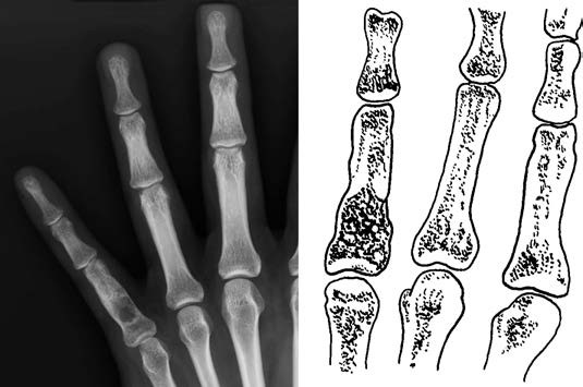
Figure 1 Enchondroma of the phalanx
Treatment
Surgical intervention is the main approach and can involve curettage or segmental resection with bone grafting. The prognosis is generally favorable.