Osteochondroma is a common benign tumor of cartilaginous origin, characterized by a bony protrusion on the surface of a bone, covered with a cartilage cap and containing a central medullary cavity. It typically occurs in adolescents and enlarges with skeletal growth, ceasing its growth once the epiphyseal plate closes. Osteochondromas are classified into two types: solitary and multiple. A solitary osteochondroma, also known as an exostosis, occurs as a single lesion. Multiple osteochondromas, referred to as multiple hereditary exostoses, often have a family history and carry a potential risk of malignant transformation. These tumors most commonly affect the metaphyseal regions of long bones, such as the distal femur, proximal tibia, and proximal humerus.
Clinical Features
Many cases are asymptomatic for a long time, with patients often seeking medical attention after discovering a bony mass incidentally. Pain may occur if the tumor compresses surrounding tissues or if inflammation develops in the bursa overlying the tumor. Physical examination often reveals a palpable mass, which may appear larger than suggested by X-ray imaging.
Radiographic Features
Osteochondromas may appear as solitary or multiple lesions. Radiographs show bony protrusions from the metaphysis extending toward soft tissues. These protrusions are connected to the normal bone via a narrow or wide stalk, with continuity of the cortical and cancellous bone between the lesion and the host bone. The medullary cavity of the lesion is contiguous with that of the bone. The cartilage cap covering the osteochondroma is not visualized on plain X-rays, but its thickness may vary. Irregular calcifications within the cartilage cap may occasionally be observed.
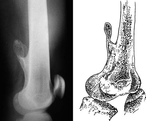
Figure 1 Osteochondroma in the distal femur
Malignant transformation of osteochondroma may result in symptoms such as pain, swelling, and the formation of soft tissue masses. Radiographically, previously stable osteochondromas may demonstrate renewed growth, bony destruction, cloud-like changes, and irregular calcifications. Solitary osteochondromas with a broad base are associated with a higher risk of recurrence.
Treatment
In general, osteochondromas do not require treatment. Surgical excision is indicated when the tumor exhibits rapid growth, causes pain, impairs joint function or adjacent bone growth, induces joint deformities, compresses nerves or blood vessels, sustains fractures, causes recurrent inflammation of an overlying bursa, or demonstrates signs suspicious for malignancy. Excision involves removing the tumor along with a margin of surrounding normal bone, including the fibrous membrane or bursa and the cartilage cap, to minimize the risk of recurrence.