Rheumatoid arthritis (RA) is a chronic inflammatory disease primarily affecting the joints, characterized by symmetrical and polyarticular onset. It is classified as an autoimmune disease. The condition frequently involves small joints such as those of the hands, wrists, and feet, with recurring episodes of joint pain and swelling that progressively worsen. Over time, it leads to joint destruction, ankylosis, and deformation.
Etiology
The exact cause of rheumatoid arthritis remains unclear, but several factors may contribute to its development:
Autoimmune Response
The human leukocyte antigen HLA-DR4 has been correlated with this disease to varying degrees. Autoimmune responses lead to synovial hyperplasia, pannus formation, inflammation, cellular infiltration, and cartilage destruction.
Infection
Certain features of the disease are similar to viral infections, and some suggest that Group A beta-hemolytic streptococcal infection might act as a trigger.
Genetic Factors
Rheumatoid arthritis has a notable genetic predisposition, with a significantly higher prevalence observed in families with a history of the disease.
Pathology
The fundamental pathological change in rheumatoid arthritis involves chronic inflammation of the joint synovium. During the early stages, the synovium becomes congested, edematous, and infiltrated by monocytes and lymphocytes, leading to the formation of granulation tissue (pannus) at the synovial edges. Over time, the pannus progressively covers the surface of the articular cartilage. In later stages, this granulation tissue undergoes fibrosis, resulting in fibrous joint stiffness, which can further evolve into bony ankylosis. Tendons and tendon sheaths around the joint are similarly invaded by granulation tissue, causing contractures and further impairing joint function.
Clinical Manifestations
Rheumatoid arthritis predominantly affects middle-aged women. Early symptoms include fatigue, myalgia, low-grade fever, and systemic manifestations, as well as recurring, symmetrical, and polyarticular arthritis. Commonly involved joints include the proximal interphalangeal joints, metacarpophalangeal joints, wrists, elbows, shoulders, knees, and metatarsophalangeal joints. The cervical spine and temporomandibular joints may also be affected, accompanied by restricted movement. Hip joint involvement is rare.
Joint Pain and Swelling
Affected joints are characterized by persistent swelling, pain, and tenderness. In the majority of cases, joint swelling is the initial symptom, caused by increased intra-articular exudate and inflammation of surrounding soft tissues. The swelling appears uniform, with spindle-shaped swelling in the proximal interphalangeal joints being a hallmark feature. The severity of joint pain is usually proportional to the degree of swelling, with more pronounced swelling associated with greater pain intensity.
Morning Stiffness
Over 95% of patients experience morning stiffness in the joints. This stiffness typically occurs after prolonged inactivity during the night and manifests as restricted movement upon waking, often lasting for more than one hour. Severe cases may involve a generalized feeling of stiffness in all joints. Physical activity or warming the joints typically alleviates or resolves the stiffness. Morning stiffness is often accompanied by sensations of coldness or numbness in the fingers and toes.
Limited Joint Mobility and Deformities
As the disease progresses, joint mobility becomes increasingly restricted. In advanced stages, various joint deformities may develop. The most common deformities in the hands include ulnar deviation of the metacarpophalangeal joints and swan-neck deformities of the fingers, where the metacarpophalangeal joints are flexed, the proximal interphalangeal joints are hyperextended, and the distal interphalangeal joints are flexed. Other deformities include knee joint valgus or varus deformities and ankylosis of the wrist and elbow joints.
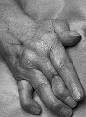
Figure 1 Ulnar deviation deformity of metacarpophalangeal joints and swan-neck deformities in fingers caused by rheumatoid arthritis
Auxiliary Examinations
Laboratory Tests
Hemoglobin levels may be reduced.
White blood cell counts are either normal or decreased, but lymphocyte counts are elevated.
Rheumatoid factor is positive in approximately 70–80% of cases.
Erythrocyte sedimentation rate (ESR) and C-reactive protein (CRP) levels are elevated.
Serum levels of immunoglobulins (IgG, IgA, and IgM) are increased.
Synovial fluid appears turbid, with reduced viscosity, poor mucin clot formation, decreased glucose levels, and negative bacterial cultures.
Imaging Studies
Early X-ray findings include soft tissue swelling around the joints, joint space widening, and localized osteoporosis near the joints.
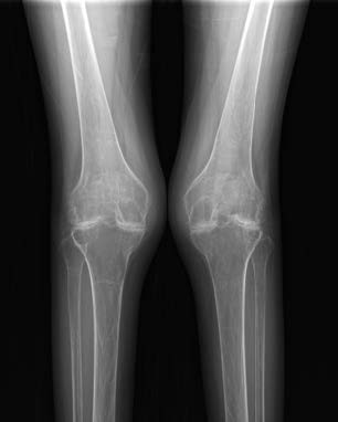
Figure 2 X-ray showing rheumatoid arthritis involvement in the knee joint
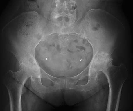
Figure 3 X-ray showing rheumatoid arthritis involvement in the hip joint
As the disease progresses, periarticular osteoporosis intensifies, joint margins become blurred, and the joint space gradually narrows.
In advanced stages, joint spaces may disappear entirely, leading to the development of bony ankylosis.
Diagnosis and Differential Diagnosis
Diagnostic Criteria
Diagnostic criteria include:
- Morning joint stiffness lasting at least 1 hour (≥6 weeks).
- Swelling in three or more joints (≥6 weeks).
- Swelling in wrist, metacarpophalangeal, or proximal interphalangeal joints (≥6 weeks).
- Symmetrical joint swelling (≥6 weeks).
- Presence of subcutaneous rheumatoid nodules.
- Clear evidence of osteoporosis or bone erosion in hand and wrist joints on X-ray.
- Positive rheumatoid factor (titer >1:32).
A diagnosis of rheumatoid arthritis can be established if four or more of these criteria are met.
Major Differential Diagnoses
Rheumatic Arthritis
Rheumatic arthritis is a systemic hypersensitivity reaction caused by infection with hemolytic streptococci, often accompanied by a history of infections such as pharyngitis or erysipelas. The onset tends to be acute and is more common in adolescents. The condition can involve the heart, leading to rheumatic heart disease, and presents with features such as fever, subcutaneous nodules, and rash. Two main characteristics are observed:
- First, affected joints exhibit marked redness, swelling, warmth, and pain, with mobility being significantly impaired. Commonly involved joints include large joints of the lower extremities, such as the knees, hips, and ankles, followed by the shoulders, elbows, and wrists, while small joints of the hands and feet are rarely affected.
- Second, the condition is characterized by migratory polyarthritis, where pain shifts between different joints but resolves within a few days.
Laboratory findings typically show an increased erythrocyte sedimentation rate (ESR) and elevated anti-streptolysin O titers, with a negative rheumatoid factor test. Recurrence after recovery is rare, and the joints are generally free from deformities.
Ankylosing Spondylitis
Ankylosing spondylitis primarily affects the spine, although peripheral joints may also be involved. The condition exhibits the following features:
- It is more common in young males.
- The primary regions affected include the sacroiliac joints and the spine, with peripheral joint involvement typically limited to large joints such as the hips, knees, and ankles. Enthesitis is frequently observed.
- Between 88% and 96% of patients test positive for HLA-B27.
- Rheumatoid factor tests are negative.
- Specific X-ray changes in the sacroiliac joints and spine are highly valuable for differential diagnosis.
Gouty Arthritis
Gouty arthritis is often observed in middle-aged and elderly men. It is typically recurrent and tends to involve the unilateral first metatarsophalangeal joint or the midfoot. Other joints, including the knees, ankles, elbows, wrists, and hands, may also be affected. Elevated serum uric acid levels are a common feature, and in some cases, tophi may form in joints or regions such as the auricles.
Osteoarthritis
Osteoarthritis is more frequently seen in middle-aged and elderly individuals. It commonly affects the knee and hip joints, although hand joints may also be involved asymmetrically. Joint swelling and morning stiffness can occur, but stiffness typically lasts less than 30 minutes. Laboratory findings are generally unremarkable, while imaging features are more distinct. A particularly notable feature is the formation of osteophytes at joint margins, which is absent in rheumatoid arthritis.
Treatment
Currently, no definitive cure exists for rheumatoid arthritis. Treatment focuses on controlling inflammation, alleviating symptoms, slowing disease progression, preserving joint function, and preventing deformities. Comprehensive treatment plans should be developed based on individual patient conditions.
Non-Pharmacological Treatment
Rest is advisable in cases of severe joint pain.
Adjustments to lifestyle are important, including smoking cessation, weight management, a balanced diet, and moderate exercise.
Joint mobility and strength training exercises are appropriate when symptoms are mild.
Pharmacological Treatment
Early initiation of treatment with conventional synthetic disease-modifying antirheumatic drugs (DMARDs) is recommended following diagnosis. Methotrexate is typically the first-line choice.
If methotrexate is contraindicated, alternatives such as leflunomide or sulfasalazine may be used.
If monotherapy proves insufficient, combination therapy with another one or two conventional synthetic DMARDs is recommended.
In cases where conventional DMARDs are ineffective, treatment may involve combining a conventional synthetic DMARD with either a biological DMARD or a targeted synthetic DMARD.
For patients with moderate or severe disease activity, low-dose glucocorticoids may be added to conventional synthetic DMARD therapy for rapid symptom control.
Surgical Treatment
Early interventions may include arthroscopic procedures such as joint debridement and synovectomy. These procedures reduce joint effusion, inhibit pannus formation, protect cartilage and subchondral bone, and improve joint function.
In advanced stages where joint function is severely impaired, total joint replacement surgery may be considered.
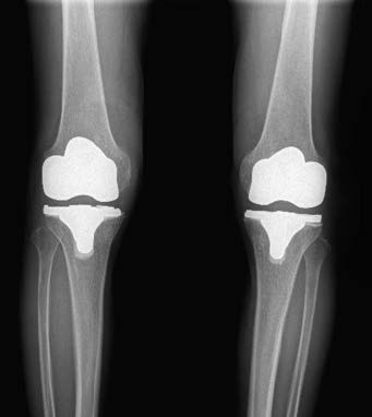
Figure 4 X-ray after total knee joint replacement in rheumatoid arthritis
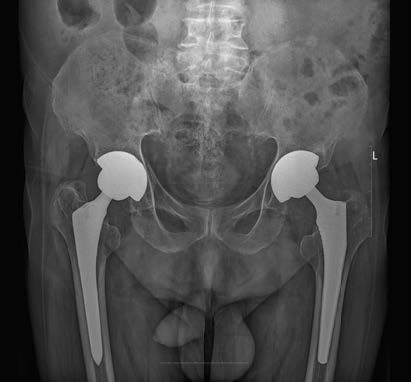
Figure 5 X-ray after total hip joint replacement in rheumatoid arthritis