Lumbar spinal stenosis is a clinical syndrome broadly defined as any form of narrowing of the spinal canal, nerve root canal, or intervertebral foramina, excluding independent clinical diseases that cause lumbar spinal canal narrowing. This narrowing can result in compression of the cauda equina or nerve roots. Based on etiology, it can be classified into congenital, developmental, and acquired spinal stenosis. Acquired causes include degenerative changes, iatrogenic factors, trauma, spondylolysis with spondylolisthesis, and others. Degenerative spinal stenosis is the most commonly observed type in clinical practice.
Etiology and Pathology
The shape of the lumbar spinal canal varies across different segments. In adults, the L1–2 segment has an oval shape, while the L3–5 segments are more often triangular or trefoil-shaped. Degenerative changes in the lumbar spine, such as intervertebral disc bulging, folding of the ligamentum flavum, osteophyte formation along the posterior vertebral edge, hypertrophy and inward displacement of the facet joints, can reduce the canal volume, leading to compression of nerve roots or the cauda equina. Additionally, venous congestion within the canal can cause neural ischemia. Prolonged compression exacerbates neural damage, although significant stenosis on imaging may exist without obvious neurological symptoms. Based on the location of the stenosis, lumbar spinal stenosis can be categorized into central canal stenosis, lateral recess stenosis, and foraminal stenosis.
Clinical Manifestations
Lumbar spinal stenosis, primarily degenerative, is most frequently seen in middle-aged and elderly populations. Patients often have a long-standing history of low back pain, later progressing to unilateral or bilateral leg pain, which worsens after standing or walking. In some cases, sensory abnormalities are also present. Activity typically exacerbates symptoms, including pain, numbness, and calf fatigue after walking certain distances, which improves with rest or squatting but recurs upon resuming activity. This is referred to as neurogenic claudication.
Physical examination often reveals pronounced symptoms and mild clinical signs. There is usually a reduction in lumbar lordosis, with normal forward flexion and restricted backward extension of the lumbar spine. Lumbar extension may provoke lumbosacral pain, or leg pain and numbness, and signs of nerve root compression may be present. Severe cases may result in compression of the cauda equina, leading to sphincter dysfunction.
Imaging Studies
X-ray imaging typically shows degenerative changes in the lumbar spine, such as osteophyte formation, reduced intervertebral space, and decreased or reversed lumbar lordosis. Lumbar CT axial images may reveal intervertebral disc bulging, facet joint hypertrophy, and inward displacement. Lumbar MRI T1-weighted images often show multiple intervertebral disc protrusions, while T2-weighted images display reduced disc signal intensity and a characteristic waist-like narrowing of the dural sac.
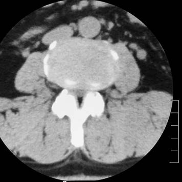
Figure 1 CT showing lumbar spinal stenosis (hypertrophy and inward displacement of the facet joints).
Differential Diagnosis
Lumbar Disc Herniation
Lumbar spinal stenosis and lumbar disc herniation present with similar symptoms, but spinal stenosis generally has fewer physical signs compared to disc herniation. The straight-leg raise test is often negative in spinal stenosis. CT imaging in spinal stenosis typically shows disc bulging rather than true herniation, along with facet joint hypertrophy and inward displacement. It is not uncommon to encounter cases where lumbar spinal stenosis coexists with lumbar disc herniation.
Facet Joint Syndrome
This condition is often observed in middle-aged women and is characterized by acute low back pain triggered by minor lumbar movements. Leg pain is usually mild or absent, and intermittent claudication is not observed. Imaging findings are generally nonspecific.
Myofascial Pain Syndrome
This condition may occur after fatigue, cooling down while sweating during activity, or an upper respiratory tract infection. Pain is commonly located in the trapezius, supraspinatus, sacrospinalis, and gluteal muscles. Imaging results are typically normal.
Treatment
Patients with mild symptoms of lumbar spinal stenosis benefit from non-surgical treatment, including bed rest to alleviate lower back pain. Physical therapy and nonsteroidal anti-inflammatory drugs (NSAIDs) can reduce symptoms. For those with persistent symptoms refractory to non-surgical treatment, severe lumbosacral pain, marked neurogenic claudication, or imaging-confirmed severe lumbar stenosis, surgical intervention is indicated. Surgical options include simple decompression surgery or decompression combined with bone graft fusion and internal fixation.