Also known as Kienböck's disease, this condition typically occurs in individuals aged 20–30 years and is classified as a form of chronic bone injury.
Etiology
The lunate is located at the center of the proximal row of carpal bones, characterized by high mobility and relatively poor stability. Its blood supply primarily relies on small vessels within the joint capsule of the radiocarpal joint and the interosseous ligaments of the carpal bones. Frequent wrist movements generate repetitive vibrations and impacts on the lunate, leading to microvascular damage and obstruction in the joint capsule and ligaments, which causes ischemia in the lunate. The increased intramedullary pressure in the ischemic lunate further impairs circulation, resulting in avascular necrosis.
Clinical Manifestations
Symptoms
The condition has a slow onset. Patients experience wrist swelling, pain, and weakness, which worsen with activity and improve with rest. As the pain intensifies, the wrist gradually becomes swollen, movement is restricted, and the patient is unable to sustain prior work activities.
Physical Examination
Mild swelling may be present on the dorsum of the wrist, and tenderness is notable in the region of the lunate. Percussion of the head of the third metacarpal bone produces pain in the lunate area. Movement in all directions of the wrist is restricted, with dorsiflexion being the most significantly limited.
X-ray Imaging
Early-stage radiographs show no abnormalities. Months later, increased bone density in the lunate becomes apparent, along with irregularities in surface smoothness, deformities, and cystic changes in the central areas of the lunate. Osteopenia may also be observed in surrounding carpal bones.
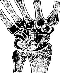
Figure 1 Avascular necrosis of the lunate
Radionuclide Bone Scintigraphy
Abnormal focal uptake in the region of the lunate may be detected in the early stages of the disease.
Treatment
At an early stage, the wrist joint can be immobilized in 20°–30° of dorsiflexion. Regular X-rays or radionuclide bone imaging are recommended during the immobilization period to monitor for recovery of lunate morphology and blood supply. Premature removal of the immobilization device risks recurrence of the condition.
For cases where the lunate is completely necrotic and deformed, lunate excision may be performed. Bone grafting or prosthesis implantation may be used to fill the defect. In cases involving physical laborers with severe osteoarthritis of the radiocarpal joint, radiocarpal joint fusion may be considered.