Anatomical Overview
The joint capsule of the knee is lax and thin, so the stability of the joint relies primarily on ligaments and muscles. The main ligaments of the knee include the medial and lateral collateral ligaments as well as the anterior and posterior cruciate ligaments.
The medial collateral ligament originates from the medial femoral epicondyle and extends downward to the medial aspect of the proximal tibia. It has superficial and deep fiber layers. The superficial layer is triangular and extends distally to the deep surface of the pes anserinus tendons. The deep layer fuses with the joint capsule and is partially connected to the medial meniscus.
The lateral collateral ligament originates from the lateral femoral epicondyle and merges distally with the tendon of the biceps femoris to form a conjoint tendon, which attaches above the fibular head. A bursa lies between the lateral collateral ligament and the lateral meniscus.
When the knee is fully extended, the collateral ligaments become taut, and adduction, abduction, and rotational movements are limited. As the knee flexes, the collateral ligaments gradually loosen, and movements such as adduction, abduction, and rotation increase.
The anterior cruciate ligament originates from the posteromedial aspect of the lateral femoral condyle and attaches anteromedially on the intercondylar eminence of the tibia. It prevents anterior translation of the tibia, especially during knee flexion and internal tibial rotation.
The posterior cruciate ligament arises from the lateral aspect of the medial femoral condyle and attaches posteroinferiorly on the tibial intercondylar eminence. It functions to prevent posterior translation of the tibia, particularly when the knee is flexed.
Mechanism of Injury and Pathology
Medial Collateral Ligament Injury
This is often caused by valgus force to the knee, such as direct impact to the lateral knee or sudden abduction and external rotation of the tibia in a semi-flexed position. This is frequently seen in sports such as football, skiing, and wrestling.
Lateral Collateral Ligament Injury
This typically results from a varus force to the knee. Due to the protective effect of the iliotibial band, isolated injuries to the lateral collateral ligament are rare and usually accompanied by fibular head fractures. In cases of high-impact trauma, injuries to the iliotibial band and the common peroneal nerve are also likely.
Anterior Cruciate Ligament Injury
This most commonly results from valgus and external rotation forces when the knee is flexed. Hyperextension of the knee may also cause anterior cruciate ligament injury. Such injuries frequently occur in sports activities and are often associated with damage to the collateral ligaments and menisci. Direct anterior impact to the posterior aspect of the proximal tibia can also cause this injury.
Posterior Cruciate Ligament Injury
This is commonly caused by a posterior force to the anterior aspect of the proximal tibia, such as falling on a flexed knee or hitting the tibial edge against a hard surface. In high-speed vehicular accidents, injuries to both anterior and posterior cruciate ligaments may occur simultaneously.
Ligament injuries may be classified as sprains (partial fiber tears), partial tears, complete tears, or combined injuries. For example, a combination of anterior cruciate ligament tear, medial collateral ligament injury, and medial meniscus tear is known as the O’Donoghue triad.
Ligament tears may occur as substance tears, avulsions at the attachment site, or avulsion fractures. Among these, substance tears tend to heal slowly and have lower tensile strength, while avulsion fractures generally heal more robustly.
Clinical Presentation and Diagnosis
A history of trauma is often reported. These injuries are more common in adolescents, particularly males, and are frequently observed among athletes. A popping sound may be heard at the time of injury, followed by severe pain that prevents continuation of activity. Swelling, restricted mobility, and muscle spasms typically develop thereafter. Palpable tenderness is usually present over the site of collateral ligament rupture.
Valgus/Varus Stress Test
This is performed with the knee fully extended and at 30° flexion to evaluate passive varus and valgus movements. Comparison with the contralateral side can reveal pain or excessive movement, suggesting collateral ligament sprain or rupture. During the acute phase, care should be taken as the test may elicit severe pain.
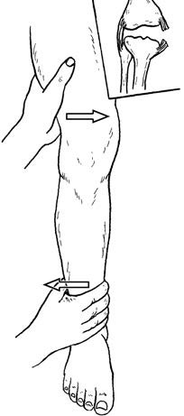
Figure 1 Valgus stress test (for medial collateral ligament assessment)
Drawer Test
With the knee flexed to 90°, the examiner stabilizes the foot and pulls the proximal tibia forward and backward. Increased anterior displacement indicates anterior cruciate ligament injury, while increased posterior displacement suggests posterior cruciate ligament injury. Comparison with the healthy side is essential.
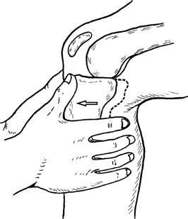
Figure 2 Positive anterior drawer test
Lachman Test
This is performed with the knee flexed 20°–30°. One hand stabilizes the distal femur while the other pulls the proximal tibia forward. The degree of tibial displacement and the firmness of the endpoint are assessed and compared with the opposite knee. This test is considered more sensitive than the anterior drawer test.
Pivot Shift Test
This is used to detect rotational instability due to anterior cruciate ligament rupture. With the patient supine and the knee extended, the examiner internally rotates the tibia while applying a valgus force at the knee. As the knee is slowly flexed to 20°–30°, a sudden shift or clunk may occur, indicating anterolateral rotational instability. This is caused by the lateral tibial plateau subluxating anteriorly and then reducing due to tension in the iliotibial band.
Imaging and Arthroscopy
Standard anteroposterior and lateral knee X-rays can reveal avulsion fractures. CT scans help define fracture extent and comminution. Stress radiographs under varus and valgus forces allow assessment of medial and lateral collateral ligament function by comparing joint space opening with the contralateral side. Stress views during anterior and posterior drawer forces assess cruciate ligament function by evaluating anterior or posterior tibial translation.
MRI provides clear visualization of ligament integrity and can detect associated injuries such as meniscal tears, cartilage damage, and occult fractures.
Arthroscopy is highly valuable for diagnosing cruciate ligament injuries. Anterior cruciate ligament damage is observed in 75% of patients with acute traumatic hemarthrosis, with two-thirds showing concurrent medial meniscus tears and one-fifth having articular cartilage defects.
Treatment
Collateral Ligament Injuries
Sprains or partial (deep layer) tears that do not significantly compromise knee stability are typically managed conservatively with bracing or plaster immobilization for 4–6 weeks. Complete tears associated with marked varus or valgus instability usually require early surgical repair or reconstruction.
Cruciate Ligament Injuries
When cruciate ligament rupture affects knee stability, arthroscopic reconstruction is often recommended. Grafts may include autologous bone–patellar tendon–bone, autologous semitendinosus–gracilis tendons, quadriceps tendon, allografts, or synthetic ligaments. For tibial spine fractures with significant displacement affecting cruciate ligament tension, surgical reduction and fixation are necessary.