Anatomical Overview
The head of the radius has an oval cross-section, with its proximal aspect forming a shallow concave articular surface that articulates with the convex head of the humerus. Together with the ulnohumeral joint, this articulation facilitates elbow flexion and extension. The ulnar side of the radial head forms the proximal radioulnar joint with the ulnar olecranon's semilunar notch, surrounded by the annular ligament. This joint, in conjunction with the distal radioulnar joint, enables forearm rotation. The radial head and neck are located within the elbow joint capsule, lacking ligamentous or tendinous attachments, which contributes to their relative instability.
Etiology and Classification
Partial dislocation of the radial head predominantly occurs in children under 5 years of age. Due to incomplete development of the radial head and aweak annular ligament, upward traction or rotation of the wrist and hand can increase intra-articular negative pressure within the elbow joint capsule, causing the weak annular ligament or portions of the joint capsule to become entrapped between the humeral head and radial head. Following removal of the traction force, the radial head fails to return to its normal anatomical position and instead displaces radially, resulting in a partial dislocation. In the vast majority of cases, the radial head undergoes radial subluxation; complete dislocation is rare, and anterior dislocation is even less common.
Clinical Presentation and Diagnosis
Children with a history of passive upward traction or injury to the wrist or hand may complain of elbow pain, restricted movement, and forearm positioning in a semiflexed and pronated position. On examination, tenderness is typically noted over the lateral aspect of the elbow, prompting a diagnosis of radial head subluxation. Radiographs often fail to demonstrate the subluxation.
Treatment
Manual reduction can be performed without anesthesia. The operator holds the child's wrist with one hand and supports the elbow with the other. The thumb is placed over the radial head, and the elbow is flexed to 90°. Gentle supination and pronation of the forearm are performed several times, with the thumb gently pressing on the radial head to achieve reduction. Successful reduction is indicated by a mild popping sound and restored normal rotation and flexion-extension of the elbow joint. No immobilization is required after reduction, but parents should be cautioned against further forceful traction to prevent recurrence.
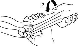
Figure 1 Reduction technique for radial head subluxation
The operator’s thumb applies pressure to the radial head while performing supination and pronation of the forearm.