Etiology
Flexion-type supracondylar humeral fractures are often caused by indirect trauma. When falling with the elbow in a flexed position and landing on the posterior aspect of the elbow, the transmitted force to the distal humerus results in a fracture.
Clinical Presentation and Diagnosis
These fractures are characterized by local swelling, pain, posterior prominence of the elbow, and subcutaneous bruising. Physical examination reveals tenderness above the elbow and the palpable fracture fragment posteriorly. X-rays typically show the presence of a fracture with characteristic displacement: the proximal fragment displaces posteriorly and inferiorly, while the distal fragment displaces anteriorly. The fracture line is oblique, angling from the anterosuperior to the posteroinferior direction. Due to the thin soft tissue covering the posterior aspect of the elbow, the sharp fracture fragment may pierce the skin, leading to an open fracture. Depending on the direction of the applied force and changes in positioning during the fall, medial or lateral displacement of the fracture may occur. However, neurovascular injuries are relatively uncommon.
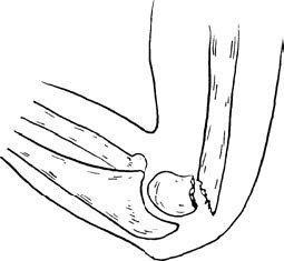
Figure 1 Typical displacement in flexion-type supracondylar humeral fractures
Treatment
The general principles of treatment are similar to those for extension-type supracondylar humeral fractures, but the direction of manual reduction differs. External fixation is performed with the elbow flexed to approximately 40°. Active elbow flexion and extension exercises can begin 4–6 weeks after reduction.
If medial or lateral displacement remains uncorrected after reduction, or if there is associated physeal injury, malunion may result in valgus or varus deformities of the elbow after fracture healing in children. Achieving anatomical reduction is recommended to prevent such complications. If anatomical reduction cannot be achieved, internal fixation with Kirschner wires may be utilized. If progressive deformity or functional impairment occurs, corrective osteotomy of the distal humerus can be performed around the age of 12–14 years. During surgery, care should be taken to avoid traction injuries to the radial and ulnar nerves. Nerve exploration may be performed before proceeding with corrective osteotomy.