Horseshoe kidney refers to a congenital anomaly where the lower poles of both kidneys are fused anterior to the abdominal aorta and inferior vena cava, forming a characteristic horseshoe shape. The incidence is approximately 25 in 10,000 individuals. In 95% of cases, the fusion occurs at the lower poles, and the isthmus typically consists of renal parenchyma, which is relatively thick and has an independent blood supply. In rare cases, the isthmus may be composed of fibrous tissue. The affected kidneys often exhibit malrotation, with the renal pelvis oriented anteriorly, the calyces directed posteriorly, and frequent variations in renal vasculature.
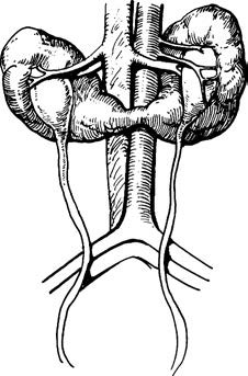
Figure 1 Horseshoe kidney
Imaging studies play a crucial role in confirming the diagnosis. Treatment is generally not required in the absence of symptoms or complications. Severe abdominal or lumbar pain, as well as gastrointestinal symptoms, may result from compression of the abdominal nerve plexus by the renal isthmus. When complications such as obstruction, stones, or infections are present, surgical interventions such as isthmus division, stone removal, or relief of obstruction may be performed.